- 2
- ,
- 2
- 4
- 6
To Quiz Yourself: Select OFF by clicking the button to hide the diagnosis & additional resources under the case.
Quick Browser: Select ON by clicking the button to hide the additional resources for faster case review.
CASE NUMBER
275


Diagnosis
Primary CNS Lymphoma
Note
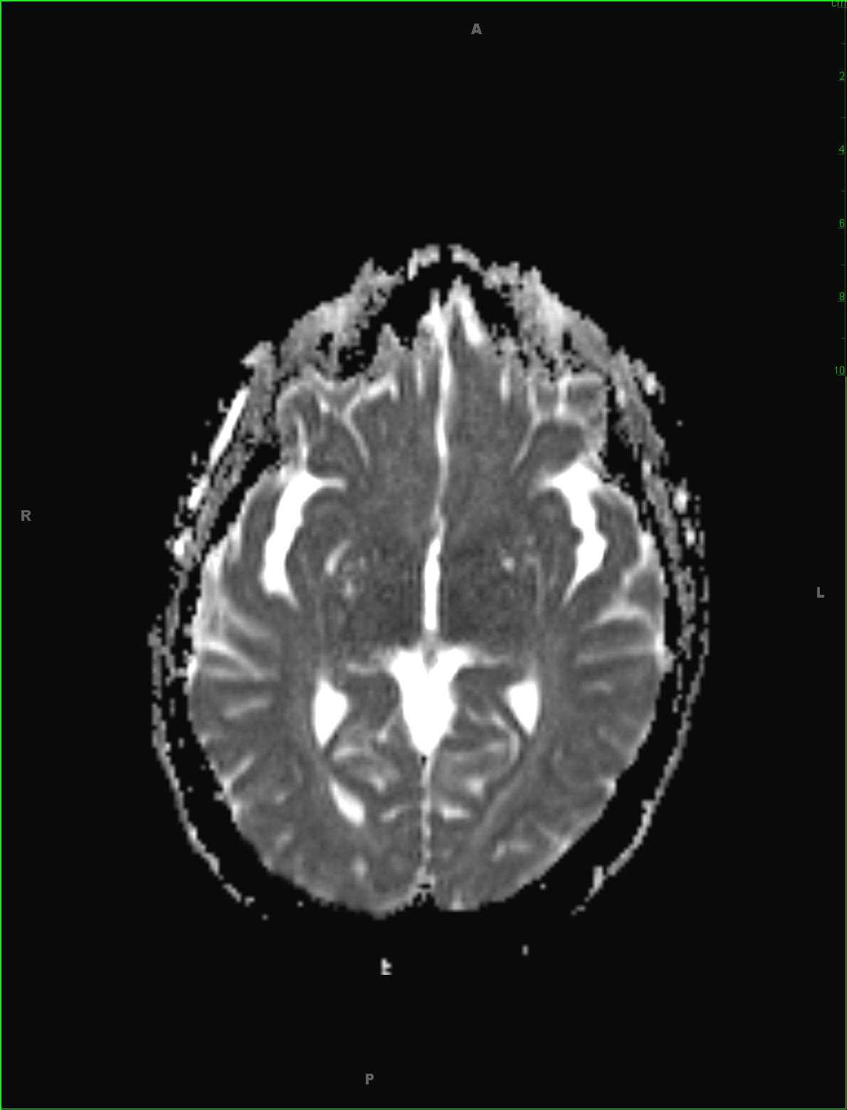
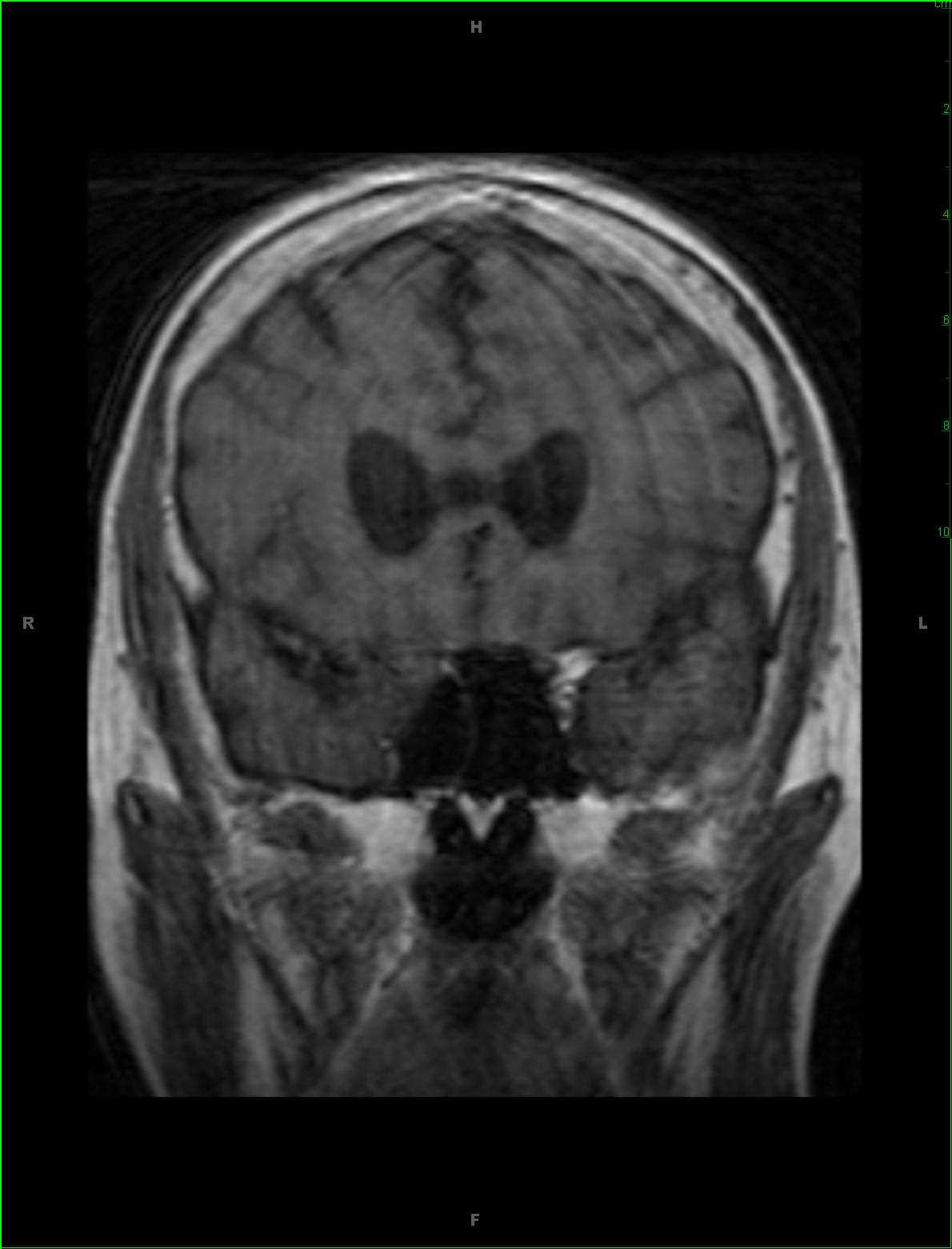
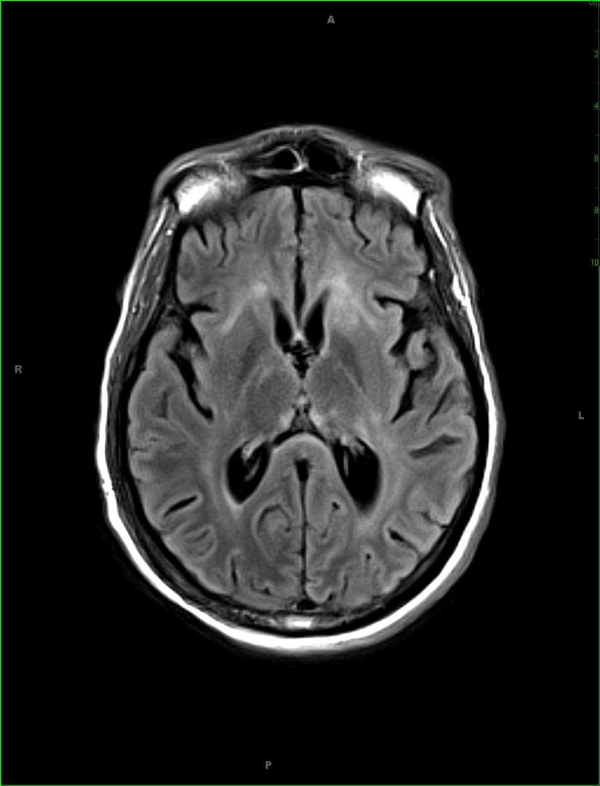
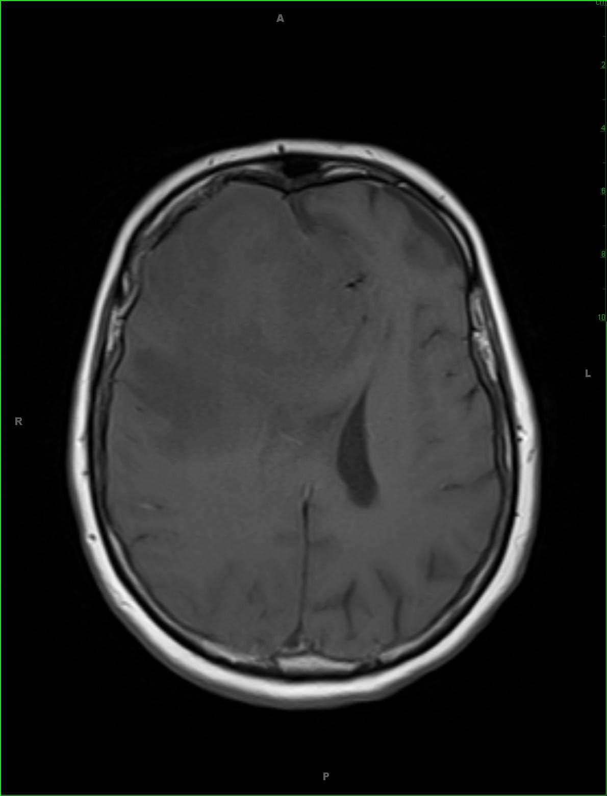
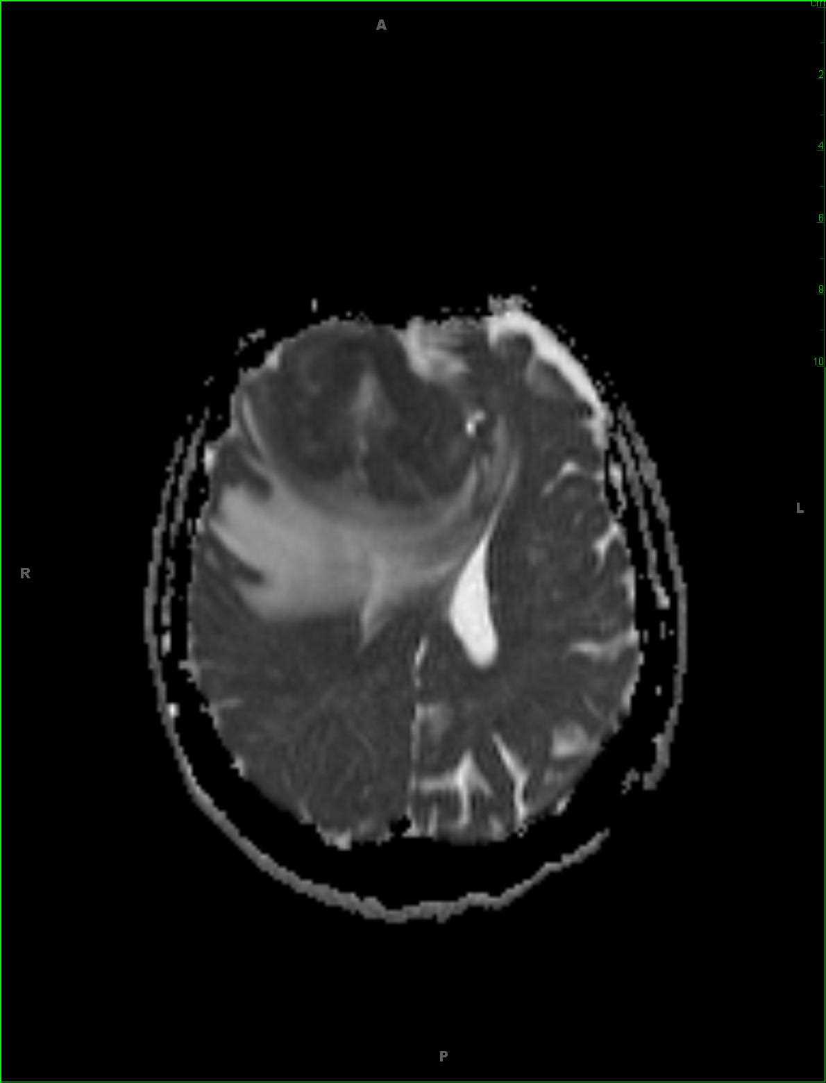
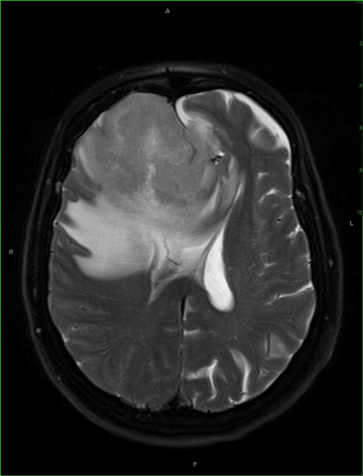
56-year-old female presented for new onset seizures. There is a partially circumscribed T1 hypointense, T2/FLAIR hyperintense lesion with involvement of both the cortical gray matter and subcortical white matter of the right frontal region. There is prominent diffusion restriction along the cortical margin and juxtacortical white matter. There is marked peripheral vasogenic edema. The lesion effaces the anterior aspect of the right lateral ventricle with lesser mass effect on the frontal horn of the left lateral ventricle. On perfusion images, there was no increased blood volume. A large confluent region of enhancement involving both the cortical gray matter and subcortical white matter is identified on the postcontrast images with the lesion extending to the overlying dural surface. On biopsy, this lesion was found to be a primary CNS lymphoma. CNS lymphoma is often associated with an immunodeficiency state, such as AIDS, organ transplantation, or collagen vascular diseases where immunomodulators are commonly used in therapy. Depending on the patients immune status and underlying chronic medical disorders, the appearance is variable with multiple necrotic masses, ventriculitis, CSF infiltration or a single necrotic mass a possibility.
Related videos to the case