- 2
- ,
- 2
- 4
- 6
To Quiz Yourself: Select OFF by clicking the button to hide the diagnosis & additional resources under the case.
Quick Browser: Select ON by clicking the button to hide the additional resources for faster case review.
CASE NUMBER
238


Diagnosis
Cortical Dysplasia
Note
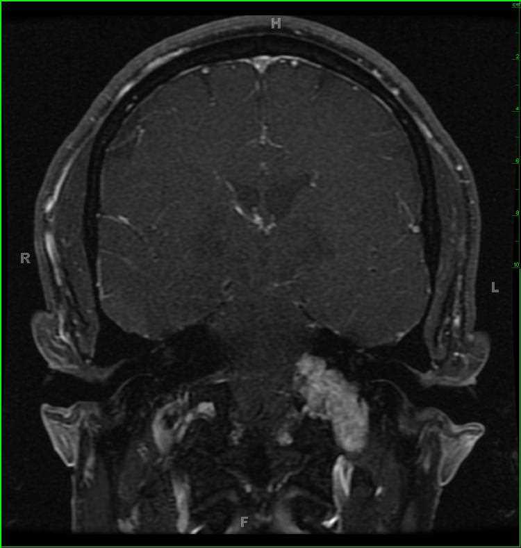
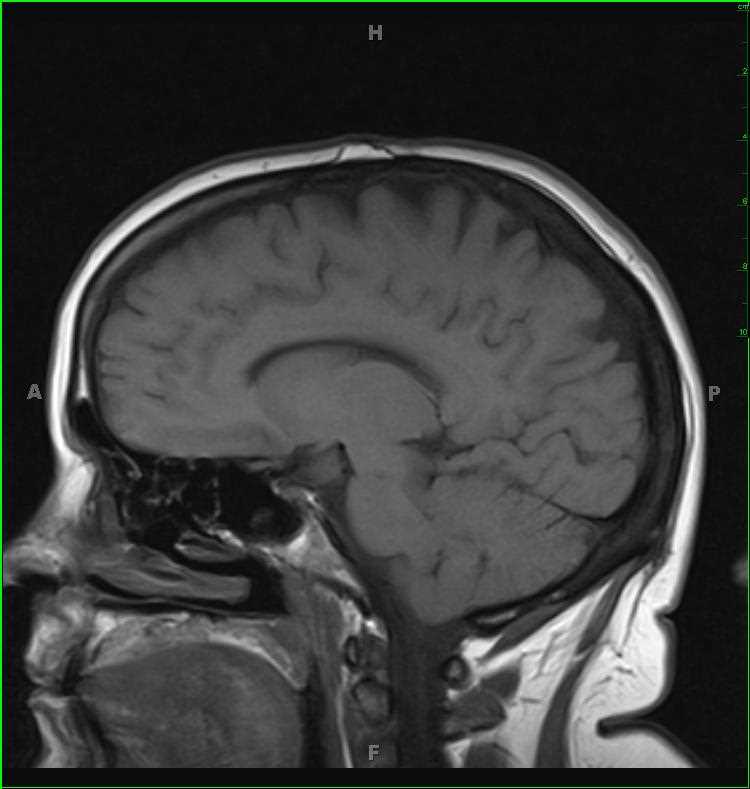
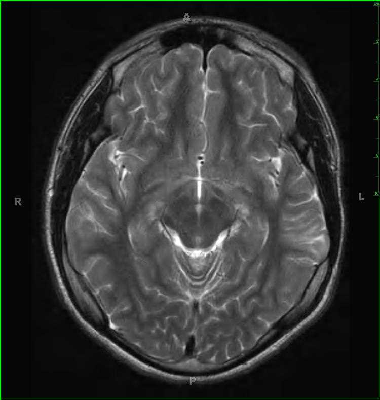
9-year-old male with history or seizures. There is a poorly circumscribed, solid and cystic, T1-hypointense, T2/FLAIR-hyperintense lesion with minimal postcontrast enhancement in the posterior aspect of the left middle temporal gyrus. The lesion involved the cortical gray matter and subcortical white matter. The differential includes focal cortical dysplasia, dysembryoplastic neuroepithelial tumor, and ganglioglioma. This lesion was resected and found to represent a focal cortical dysplasia type 1b. Focal cortical dysplasia (FCD) can be divided into two categories, FCD type I (non-Taylor dysplasia) and FCD type II (Taylor dysplasia). FCD type Ia results from dyslamination and mild malformation of cortical development. FCD type Ib results from isolated architectural abnormalities and cytoarchitectural dysplasia. FCD type II can further be subdivided into type IIa, no balloon cells, and
type IIb, balloon cells present.
Related videos to the case