- 2
- ,
- 2
- 4
- 6
To Quiz Yourself: Select OFF by clicking the button to hide the diagnosis & additional resources under the case.
Quick Browser: Select ON by clicking the button to hide the additional resources for faster case review.
CASE NUMBER
236


Diagnosis
Jugular Foramen Schwannoma, left
Note
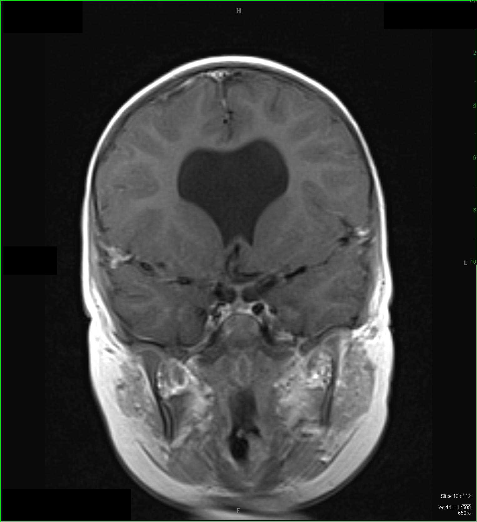
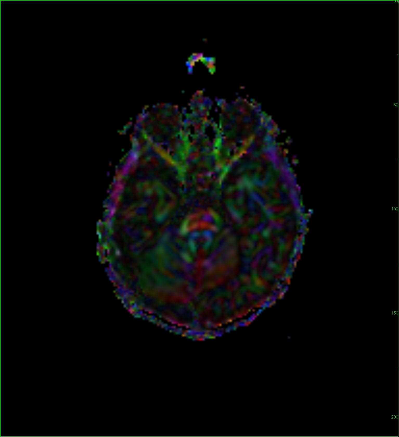
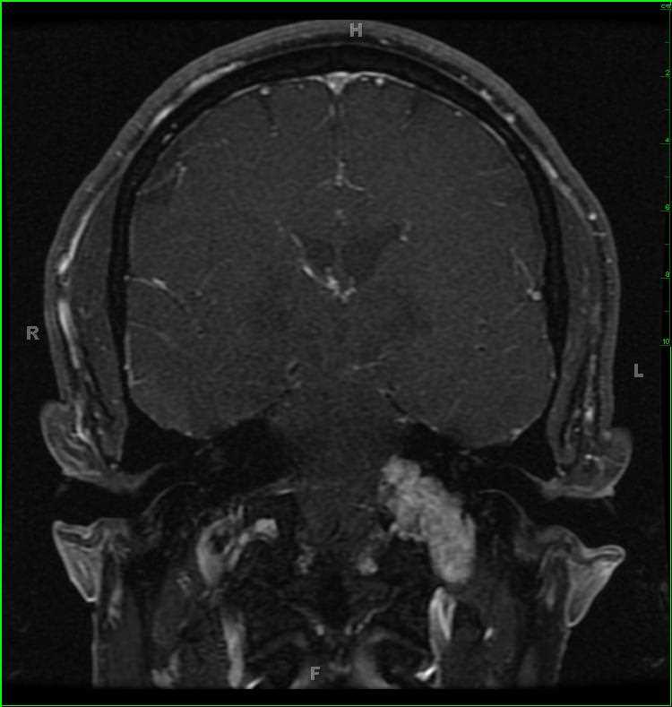
37-year-old female with history of left-sided hearing loss and vertigo. There is a T1-hypointense, multicystic, T2/STIR-hyperintense, heterogeneously enhancing mass centered within the left jugular foramen. The mass extends from the upper carotid sheath at its inferiormost extent to the left cerebellopontine angle in its rostralmost extent. There are numerous fluid/debris levels on the axial T2-weighted image which drop out on the axial gradient echo image, compatible with hemorrhage at those sites. The imaging findings are most consistent with a jugular foramen schwannoma. Jugular foramen schwannoma are benign tumors of differentiated Schwann cells involving the hypoglossal, vagus, or accessory cranial nerves. Typical age at presentation is 45. Sensorineural hearing loss is present in 90%. Complete surgical removal is the goal.
Related videos to the case