- 2
- ,
- 2
- 4
- 6
To Quiz Yourself: Select OFF by clicking the button to hide the diagnosis & additional resources under the case.
Quick Browser: Select ON by clicking the button to hide the additional resources for faster case review.
CASE NUMBER
229


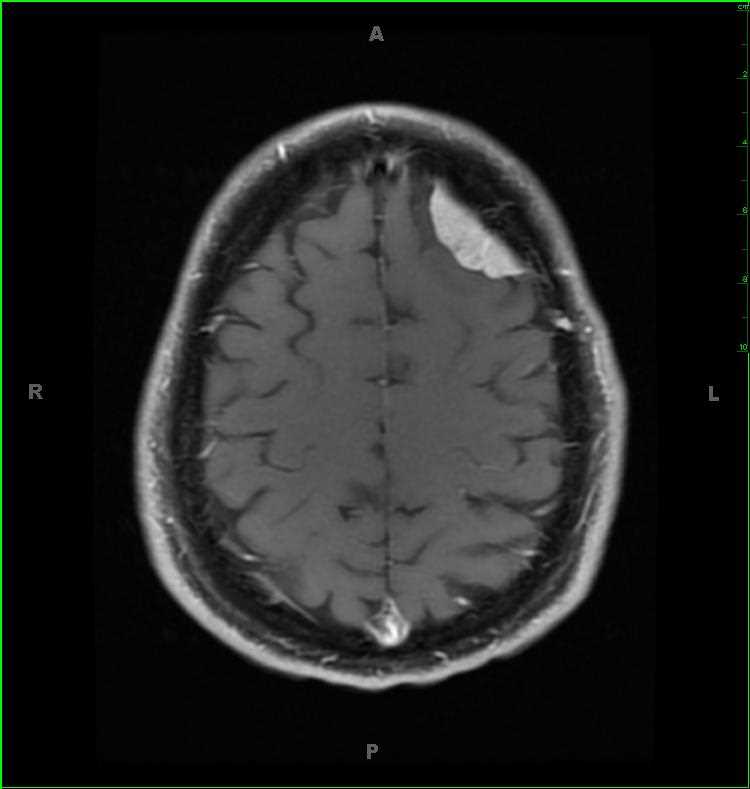
Diagnosis
Meningioma, left frontal
Note
Otherwise asymptomatic 48-year-old male, there is a well circumscribed, extra-axial, dural based, T1-hypointense, T2/FLAIR-mildly hyperintense, enhancing mass compatible with a meningioma. There is hypercellularity on the diffusion weighted images, and the lesion is partially calcified. There is partially effacement of the the underlying cerebral cortical gyri without perilesional edema. Meningioma are the most common extra-axial masses of the central nervous system. They are typically benign, and complete surgical resection is usually curative. Meningiomas are up to two times more common in females intracranially. They can be seen in the setting of NF-2.
Related videos to the case