- 2
- ,
- 2
- 4
- 6
To Quiz Yourself: Select OFF by clicking the button to hide the diagnosis & additional resources under the case.
Quick Browser: Select ON by clicking the button to hide the additional resources for faster case review.
CASE NUMBER
224


Diagnosis
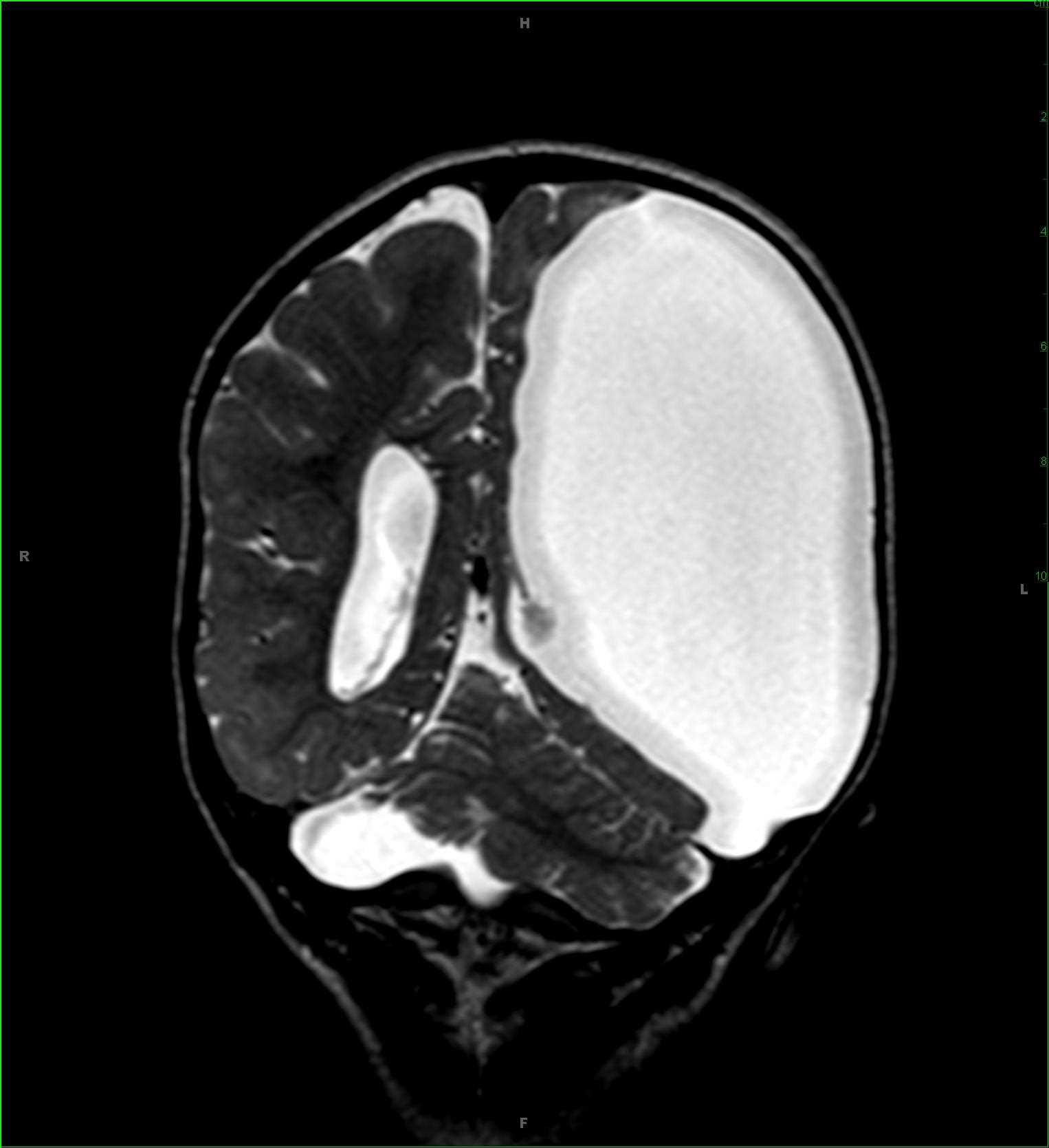
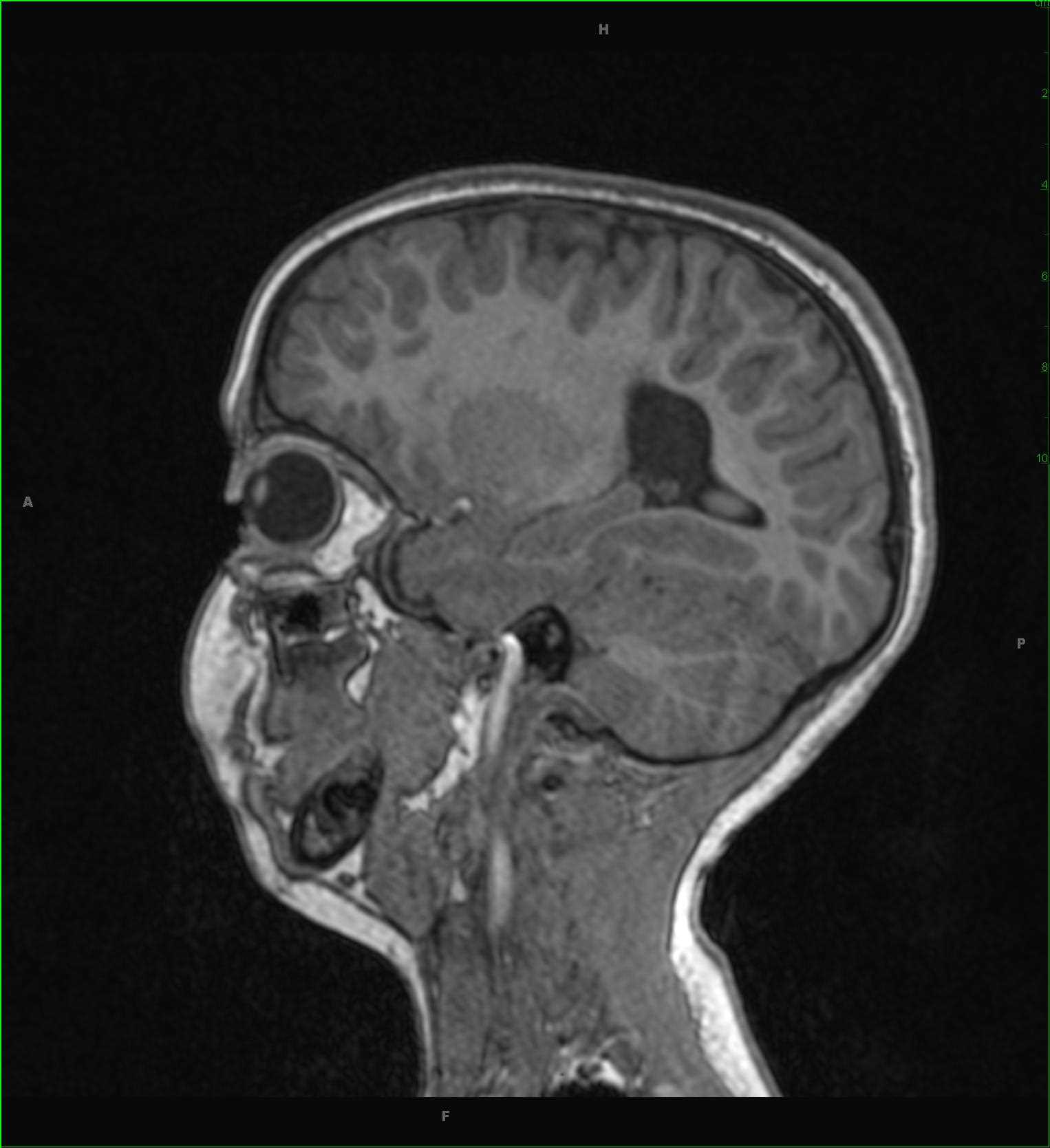
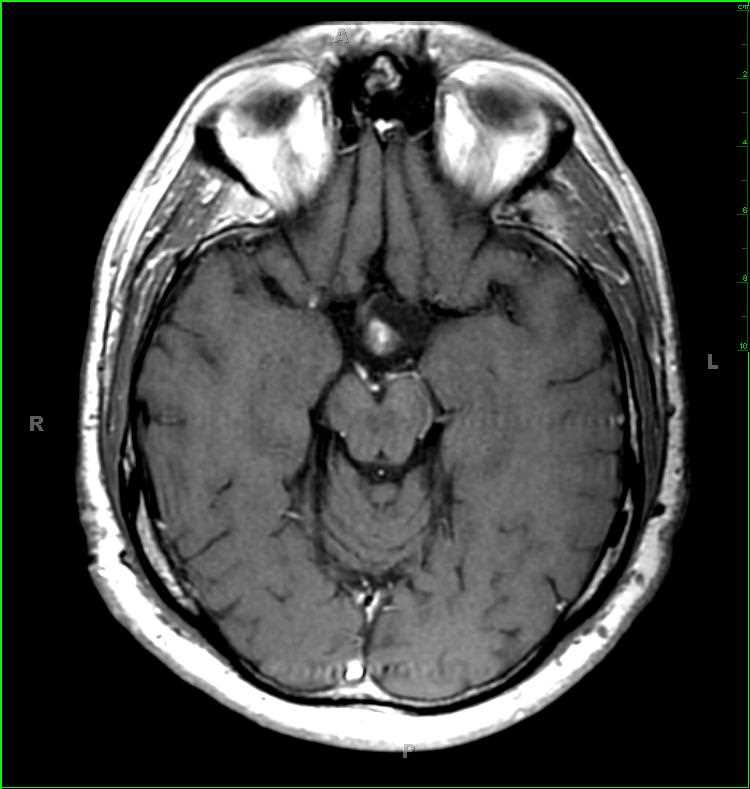
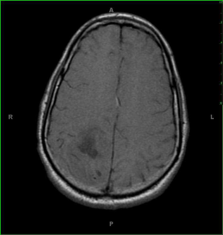
Oligodendroglioma
Note
52-year-old male with new onset seizures. Within the right parietal lobe, with minimal extension into the posterior right frontal lobe, there is a cortical and subcortical infiltrative solid and cystic T1-hypointense, non-enhancing mass. The solid components of the mass are T2/FLAIR-hyperintense, while the cystic components are T2-hyper- and FLAIR-hypointense. The involved gyri are enlarged resulting in effacement of the surrounding cerebral cortical sulci. There are components of the mass which are hypercellular on the diffusion weighted image. There are no suspicious abnormalities on the axial gradient echo image. The differential includes oligodendroglioma, astrocytoma, and ganglioglioma. This was a pathologically proven oligodendroglioma. Oligodendrogliomas account for 5-25% of all gliomas and 5-10% of all intracranial neoplasms. They typically occur in middle age, between the 4th and 5th decades. With the prominent cortical involvement, seizures are the most common clinical symptom.
Related videos to the case