- 2
- ,
- 2
- 4
- 6
To Quiz Yourself: Select OFF by clicking the button to hide the diagnosis & additional resources under the case.
Quick Browser: Select ON by clicking the button to hide the additional resources for faster case review.
CASE NUMBER
225


Diagnosis
Hypothalamic Glioma in Neurofibromatosis Type I
Note
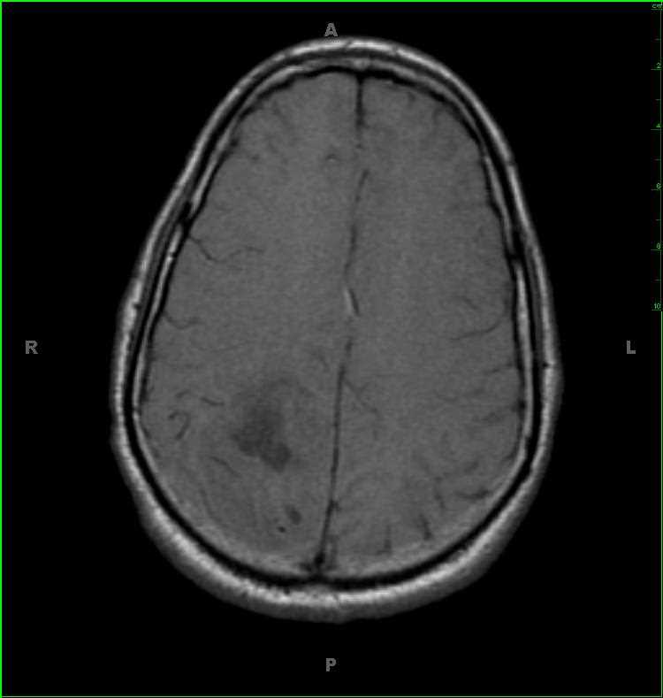
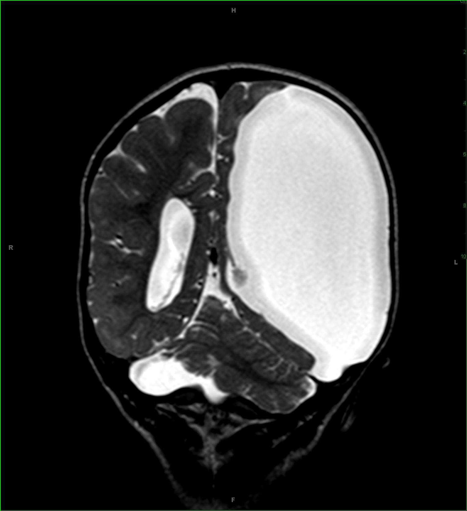
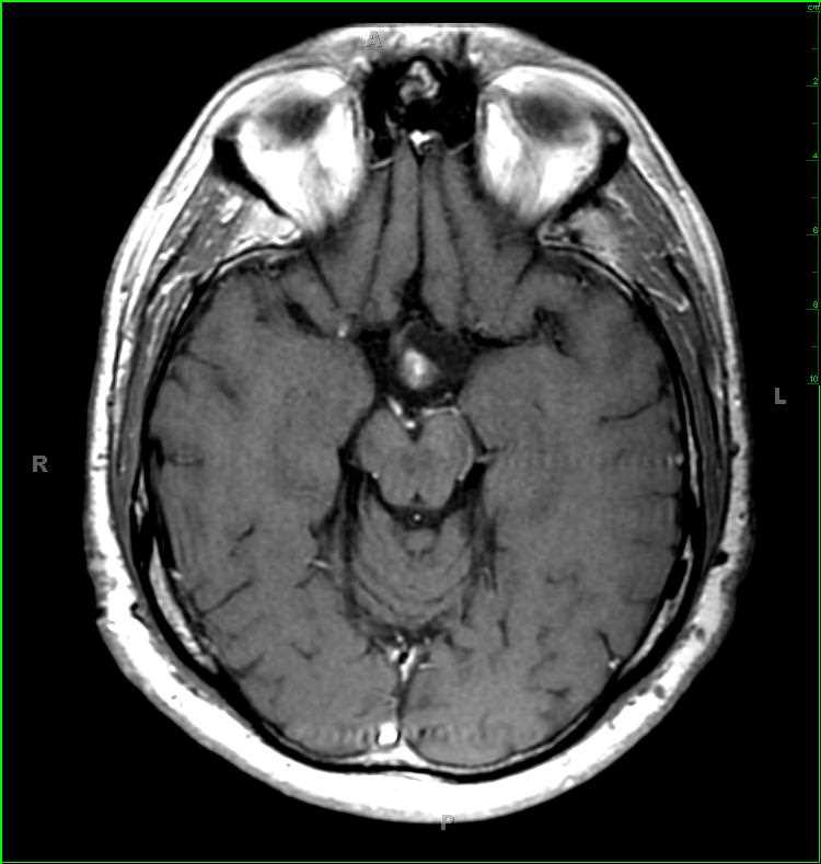
36-year-old female with history of neurofibromatosis type 1 and hypothalamic glioma. Involving the inferior most aspects of the right and left hypothalami and extending to involve the infindibulum, there is a T2/FLAIR and T1-centrally hypointense, peripherally isointense bulbous mass with heterogeneous central enhancement. There is a small exophytic cystic component of the lesion which extends anteriorly and towards the left. The lesion is distinct form the posteriormost aspect of the optic chiasm. The last image demonstrates a subcutaneous neurofibroma in the left suboccipital region. Hypothalamic gliomas may or may not extend to involve the optic chiasm, and tend to occur in the setting of NF-1. Between 20-50% of those with hypothalamic-chiasmatic glioma have a family history of NF-1. Hypothalamic-chiasmatic gliomas in the setting of NF-1 tend to have a more indolent course, and may occasionally spontaneously regress. On histologic section, the lesions are juvenile pilocytic astrocytomas. Cystic change and enhancement are common.
Related videos to the case