- 2
- ,
- 2
- 4
- 6
To Quiz Yourself: Select OFF by clicking the button to hide the diagnosis & additional resources under the case.
Quick Browser: Select ON by clicking the button to hide the additional resources for faster case review.
CASE NUMBER
222


Diagnosis
Periventricular White Matter Injury of Prematurity
Note
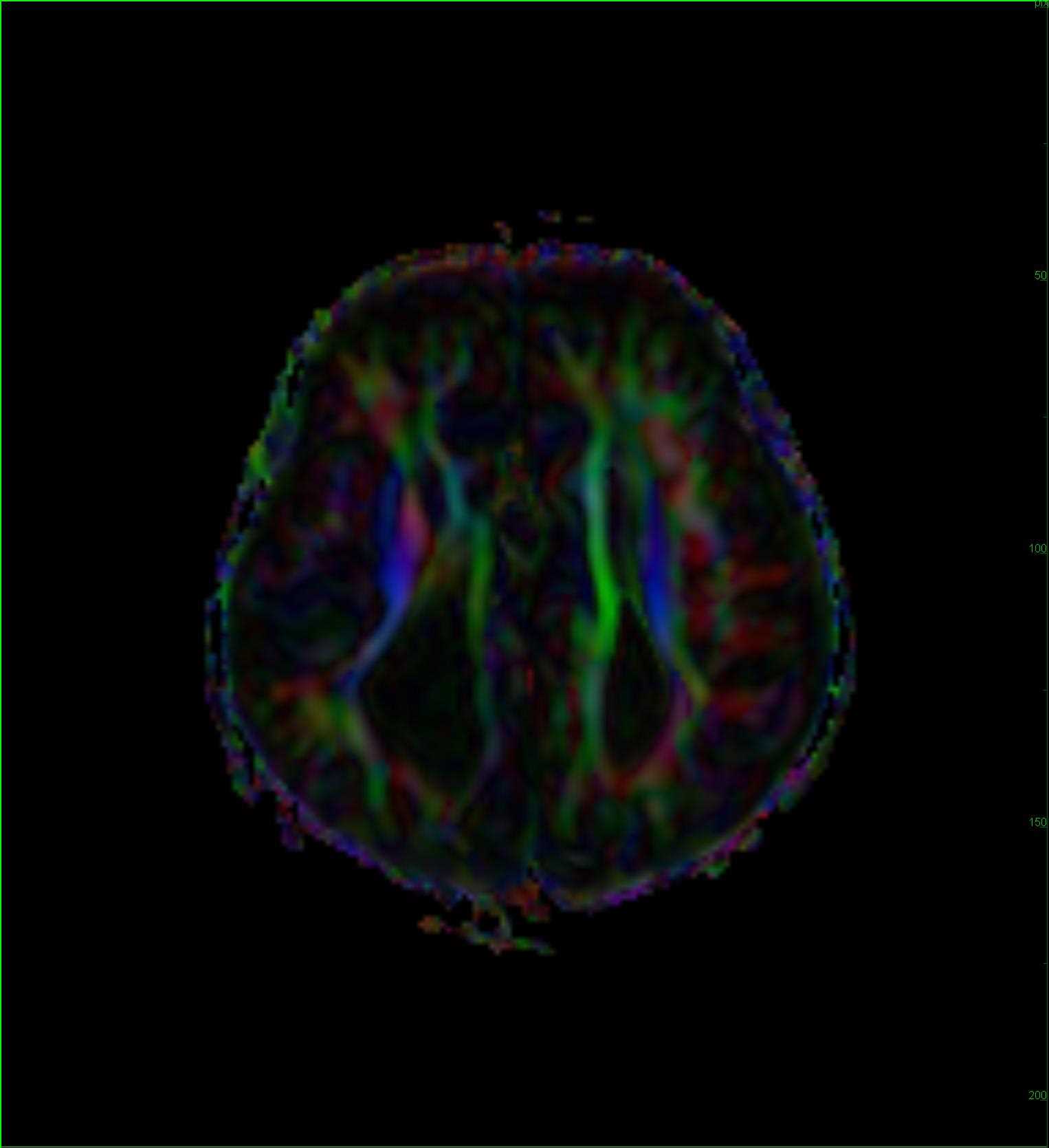
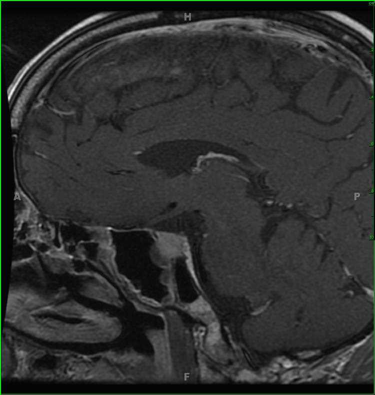
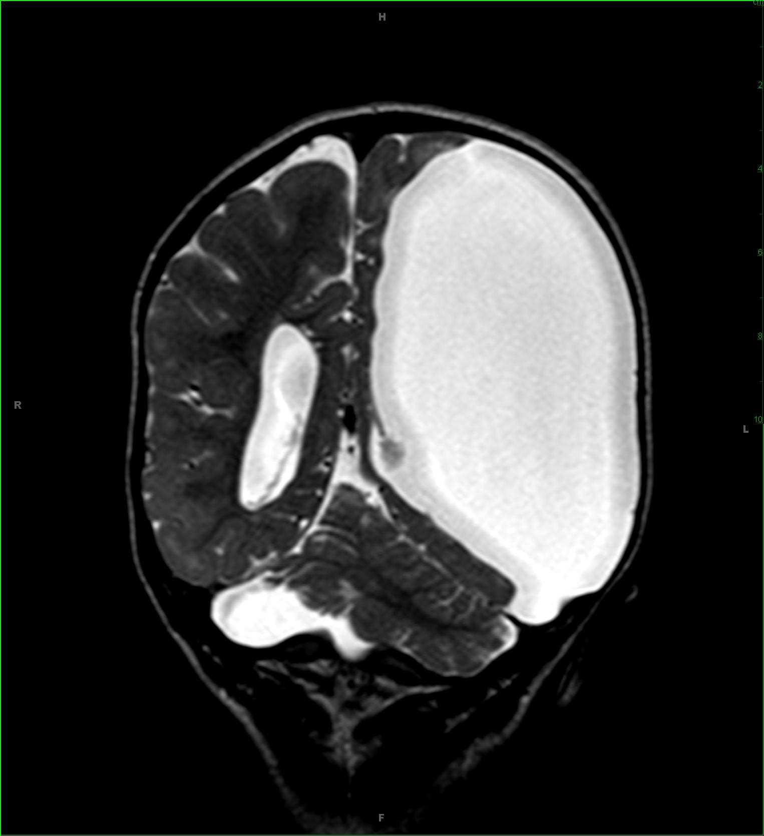
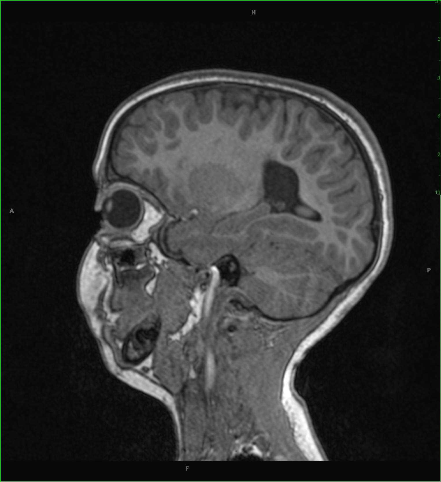
Otherwise asymptomatic 5-year-old male who was born at 28-weeks gestation. Profound periventricular white matter volume loss worse in the periatrial regions of the right greater than left cerebral hemispheres. There is ex vacuo dilitation of the lateral ventricles at those sites. There are no suspicious abnormalities on the diffusion weighted images. These findings are compatible with periventricular white matter injury of prematurity. In addition to volume loss, other imaging features include cystic cavitations and periventricular cyst formation. Periventricular white matter injury of prematurity is more common in patients born before 33 weeks gestation and weighing less than 1500 grams at birth. The disease likely results from hypoxic ischemic injury to the watershed areas, which are located in the periventricular regions in infants. Early on, periventricular white matter necrosis is seen, with cyst formation in the subacute phase, and parenchymal volume loss with ex vacuo dilitation of the ventricles in the late phase.
Related videos to the case