- 2
- ,
- 2
- 4
- 6
To Quiz Yourself: Select OFF by clicking the button to hide the diagnosis & additional resources under the case.
Quick Browser: Select ON by clicking the button to hide the additional resources for faster case review.
CASE NUMBER
218


Diagnosis
Tuberous Sclerosis
Note
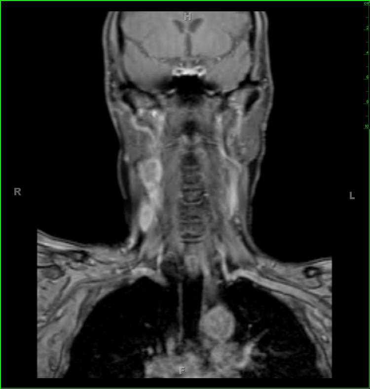
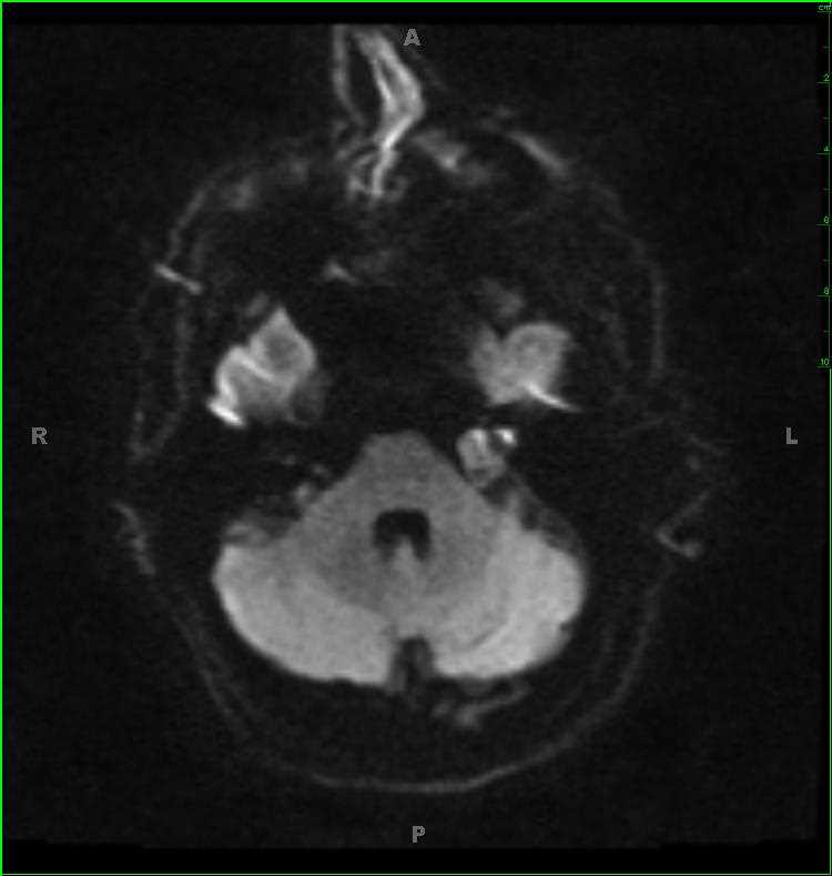
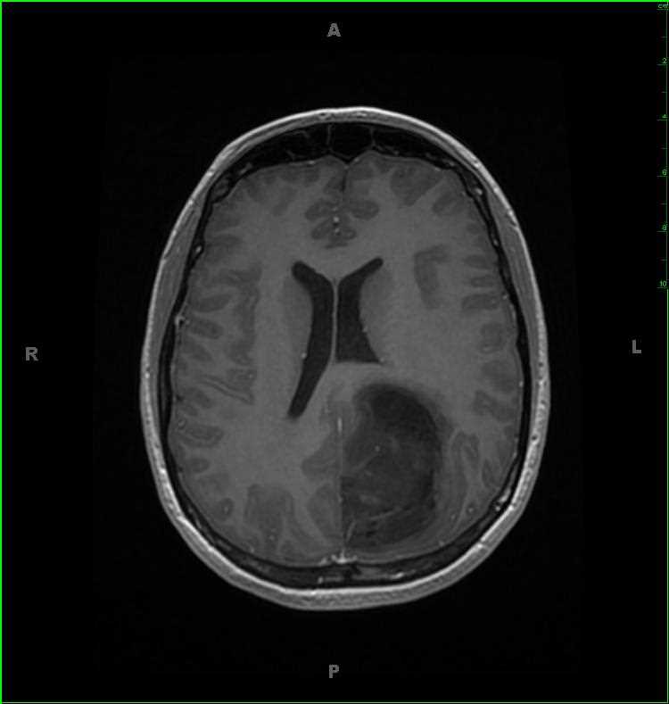
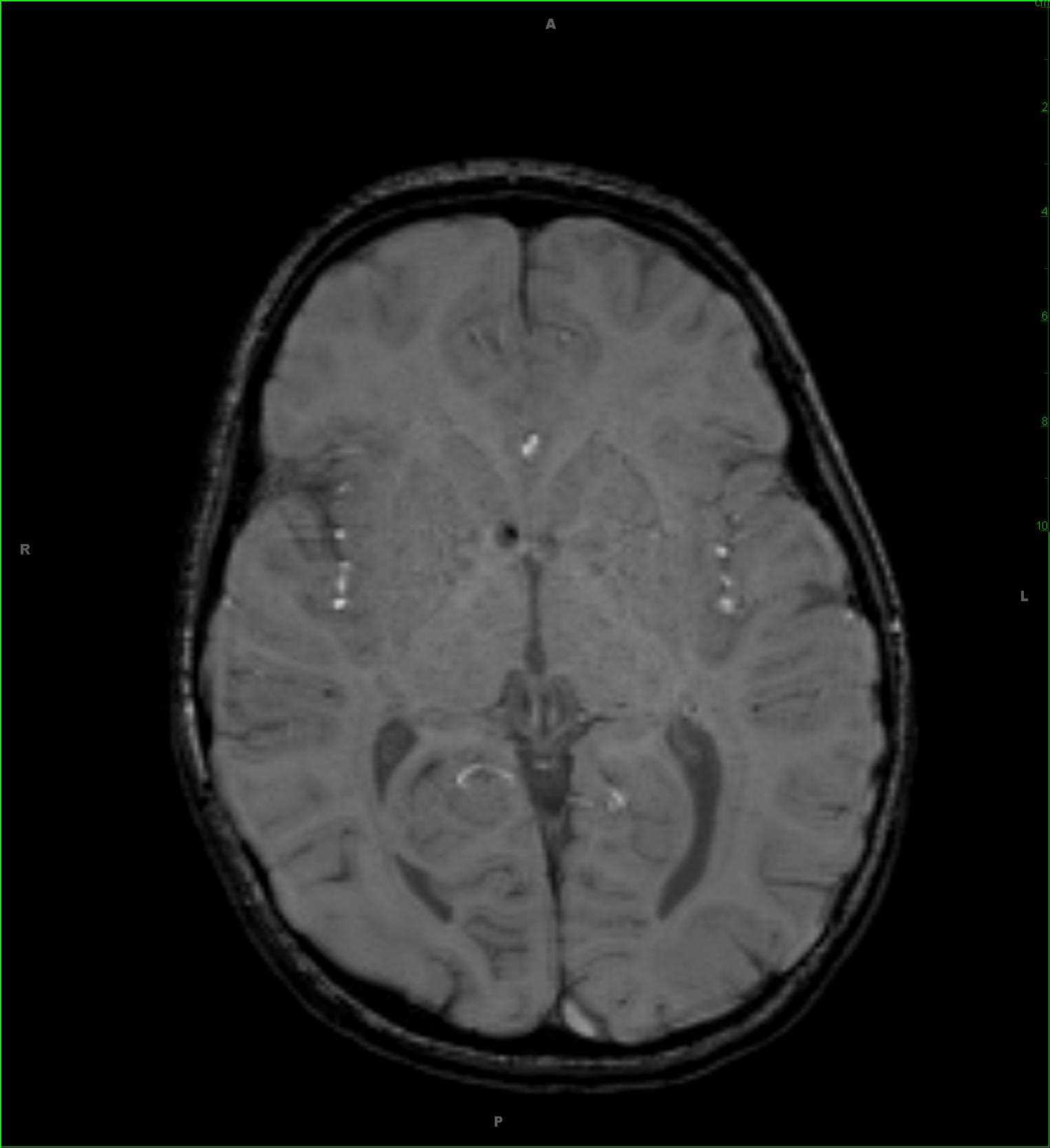
15-year-old male with history of tuberous sclerosis. On the MR images of the brain, there are numerous T2/FLAIR-hyperintense bands within the periventricular and deep white matter of the supratentorial compartment compatible with radial bands. In addition, there is a cortical/subcortical tuber in the left superior frontal gyrus region with an addition tuber in the right frontal convexity region. There are small subependymal hamartomas along the lateral margins of the right and left lateral ventricles. The final image demonstrates a focus of susceptibility at the inferior most aspect of the foramen of Monro on the right. This nodule enhanced and was T2/FLAIR hyperintense, compatible with a small subependymal giant cell astrocytoma at that site. Tuberous sclerosis is also known ad Bourneville disease, and falls into the phakomatoses. The disease results in the development of numerous benign tumors of the embryonic ectoderm. Gene mutations are on chromosome 9 (TSC1) or 16 (TSC2). Imaging findings, outside of the brain include: renal angiomylipomas, cardiac rhabdomyomas, retinal phakomas, renal cysts, renal cell carcinoma and oncocytoma and thoracic and retroperitoneal lymphangiomatosis.
Related videos to the case


THIS IS CASE
218
OF
373