- 2
- ,
- 3
- 8
- 1
To Quiz Yourself: Select OFF by clicking the button to hide the diagnosis & additional resources under the case.
Quick Browser: Select ON by clicking the button to hide the additional resources for faster case review.
CASE NUMBER
220
Diagnosis
Anaplastic Astrocytoma
Note
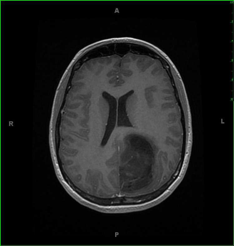
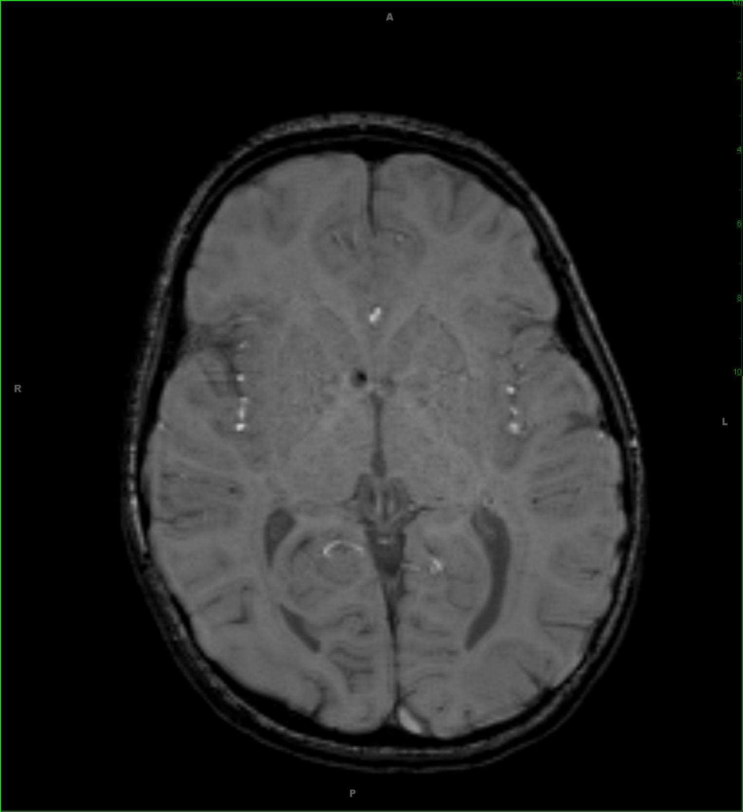
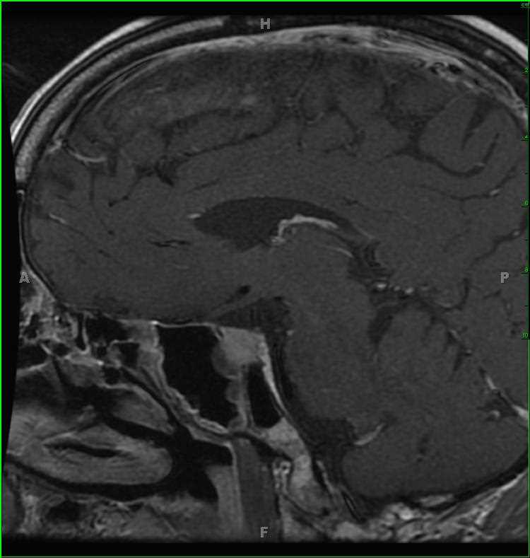
31-year-old female with a history of anaplastic astrocytoma. There is a solid and cystic mass within the left parietal lobe with a few regions of T1-hyperintense signal compatible with calcification and/or mineralization. The lesion is T2/FLAIR-hyperintense, does not restrict diffusion, and demonstrates a few faint regions of contrast enhancement. On the axial gradient echo weighted images, there are a few calcifications/mineralizations or areas of hemosiderin deposition at its posteriormost aspect. Anaplastic astrocytoma is considered a WHO Grade III tumor. These lesions usually occur in adulthood, with peak incidence between 40 and 50 years. Whereas diffuse low grade astrocytoma will not demonstrate enhancement, anaplastic astrocytoma will lack regions of frank necrosis, a finding seen in glioblastoma, WHO Grade IV tumors.
THIS IS CASE
220
OF
396


