- 2
- ,
- 2
- 4
- 6
To Quiz Yourself: Select OFF by clicking the button to hide the diagnosis & additional resources under the case.
Quick Browser: Select ON by clicking the button to hide the additional resources for faster case review.
CASE NUMBER
202


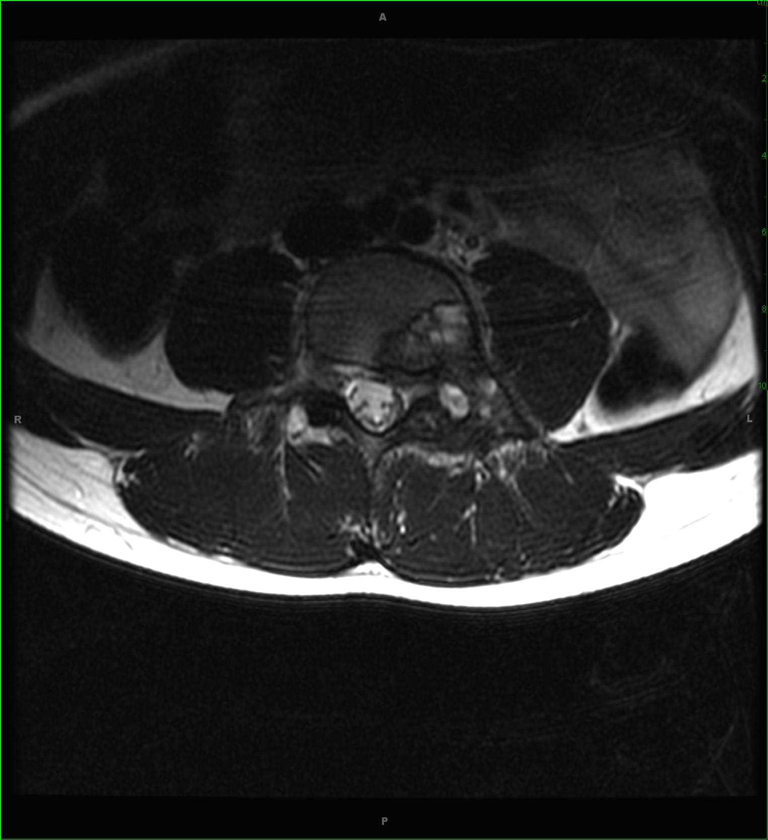
Diagnosis
Aneurysmal Bone Cyst, secondary to osteoblastoma
Note
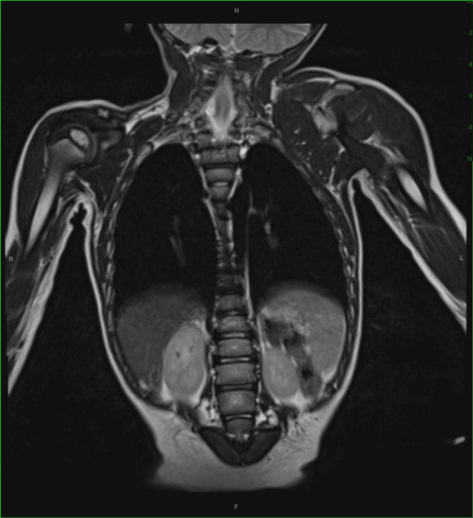
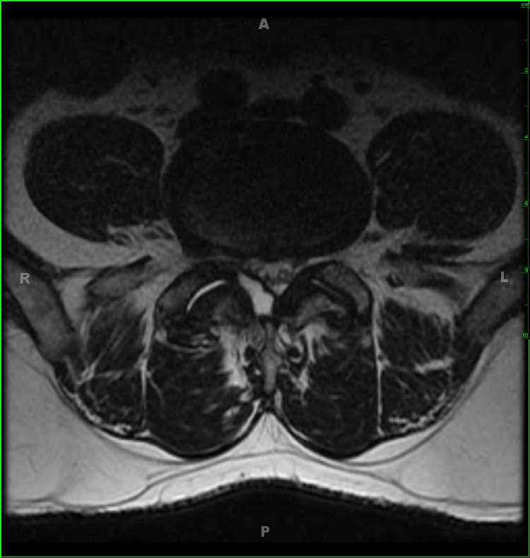
12-year-old male with left lower back pain. There is an expansile T1-isointense mass centered on the left pedicle of vertebra L4. There is isointense soft tissue which extends into the epidural space. There is also invasion of the left posterior aspect of the L4 vertebral body, left L4 lamina, and transverse process of L4 on the left. On the STIR-weighted images, the lesion is heterogeneous in signal intensity, with regions of hypo-, iso-, and hyperintense signal. On the axial STIR-weighted images, there are subtle dependent fluid/debris levels. There is also evidence of hemosiderin deposition along the margins of the lesion. The differential includes primary aneurysmal bone cyst (ABC), telangiectatic osteosarcoma, and secondary aneurysmal bone cyst. Secondary causes include giant cell tumor, chondroblastoma, osteoblastoma, and fibroxanthoma. This is an ABC in a giant osteoblastoma. ABCs represent variable-sized hemorrhage filled spaces separated by connective tissue. Up to 1/3 of ABCs are of the secondary type. The most common locations include long bones, spine/sacrum, craniofacial and epiphyses.
Related videos to the case