- 2
- ,
- 2
- 4
- 6
To Quiz Yourself: Select OFF by clicking the button to hide the diagnosis & additional resources under the case.
Quick Browser: Select ON by clicking the button to hide the additional resources for faster case review.
CASE NUMBER
200


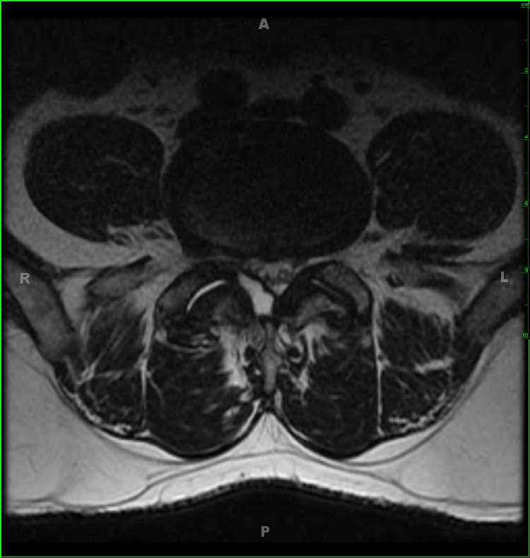
Diagnosis
Lumbar Spine Synovial Cyst
Note
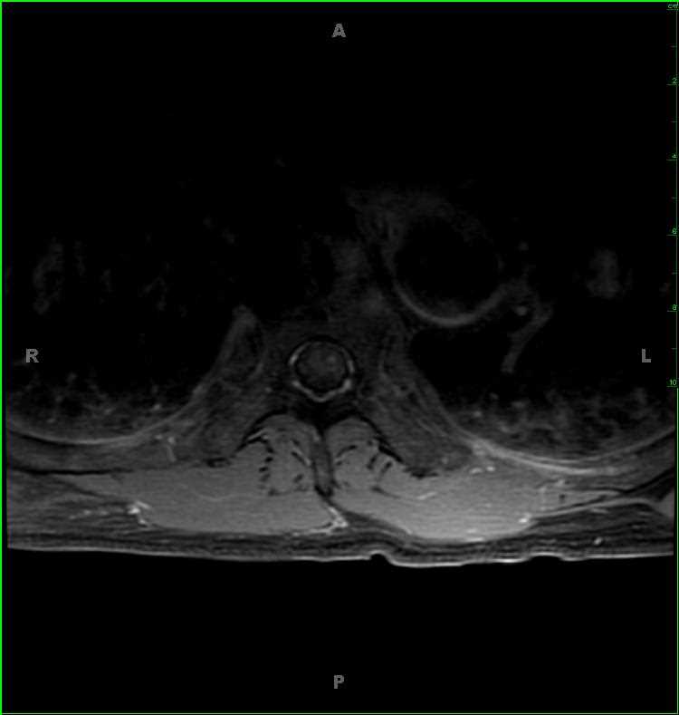
62-year-old male with worsening right-sided back pain over a period of two months. There is a well circumscribed T1-hypointense, T2/STIR-hyperintense lesion arising from the facet joint at L4-L5 on the right and extending under the ligamentum flavum. There is mild-moderate bilateral facet arthropathy. The imaging findings are most compatible with a right-sided synovial cyst which contacts the L5 and S1 nerve roots on the right. Spinal synovial cysts are collections of synovial fluid contained by a cuboid or pseudostratified columnar epithelium. The cyst may contain old hemorrhage or proteinaceous debris, which can alter its signal characteristics. Most cases are asymptomatic, but slow growth over time can result in progressive back pain. They tend to occur at the L3-L4 and L4-L5 disc levels.
Related videos to the case