- 2
- ,
- 2
- 4
- 6
To Quiz Yourself: Select OFF by clicking the button to hide the diagnosis & additional resources under the case.
Quick Browser: Select ON by clicking the button to hide the additional resources for faster case review.
CASE NUMBER
184


Diagnosis
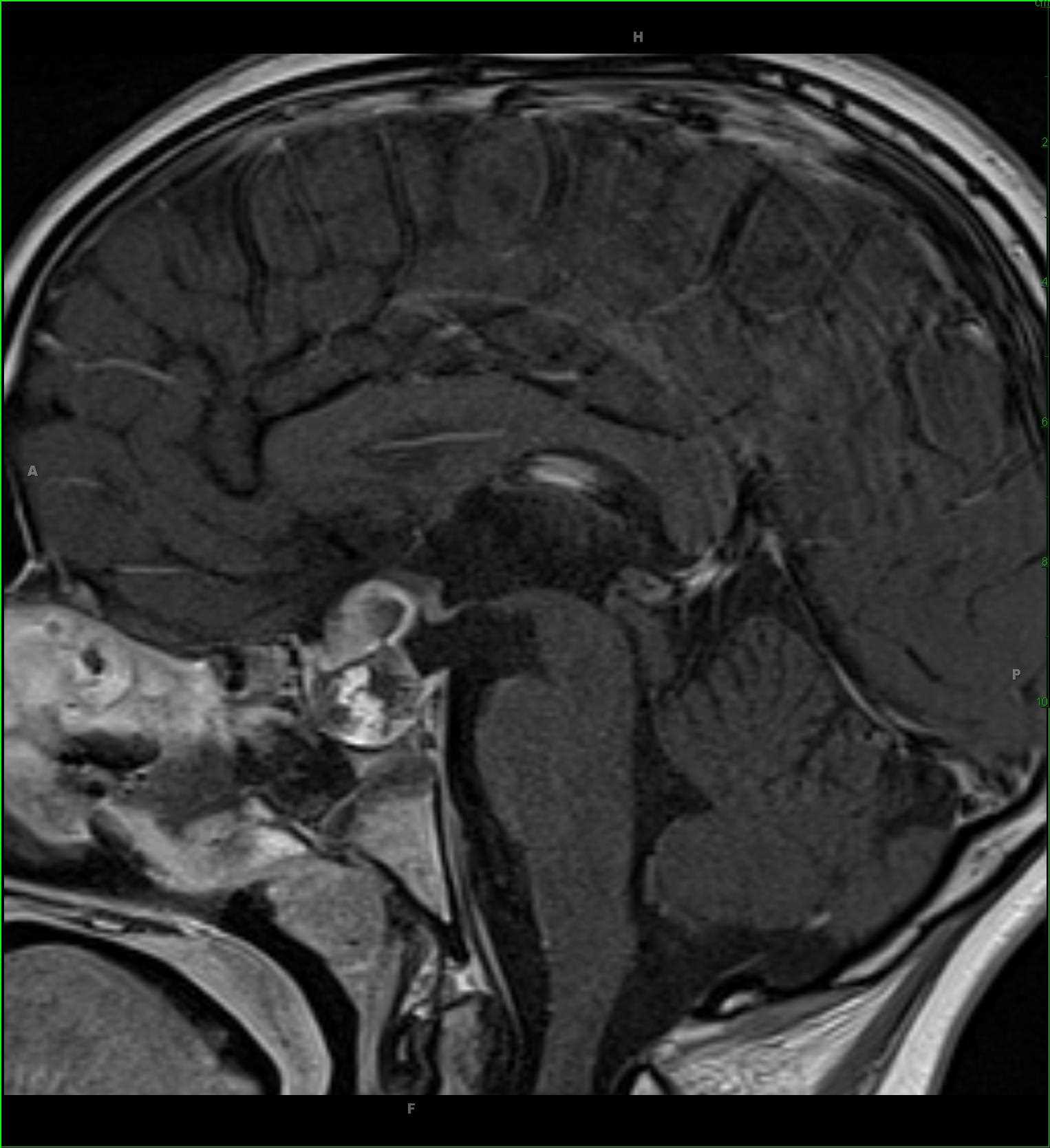
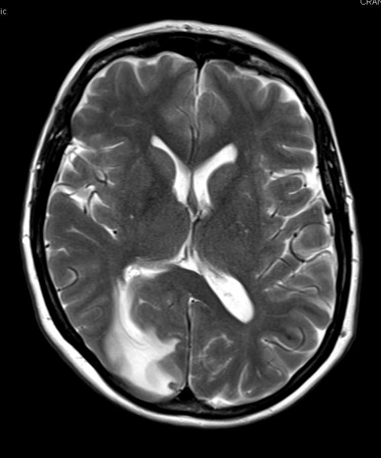
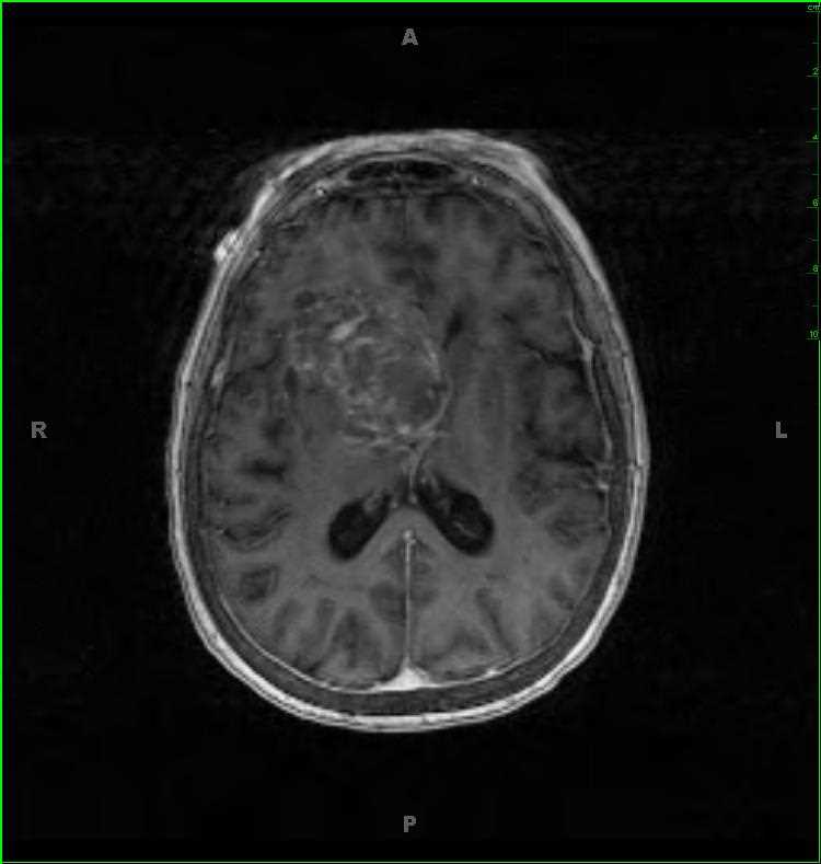
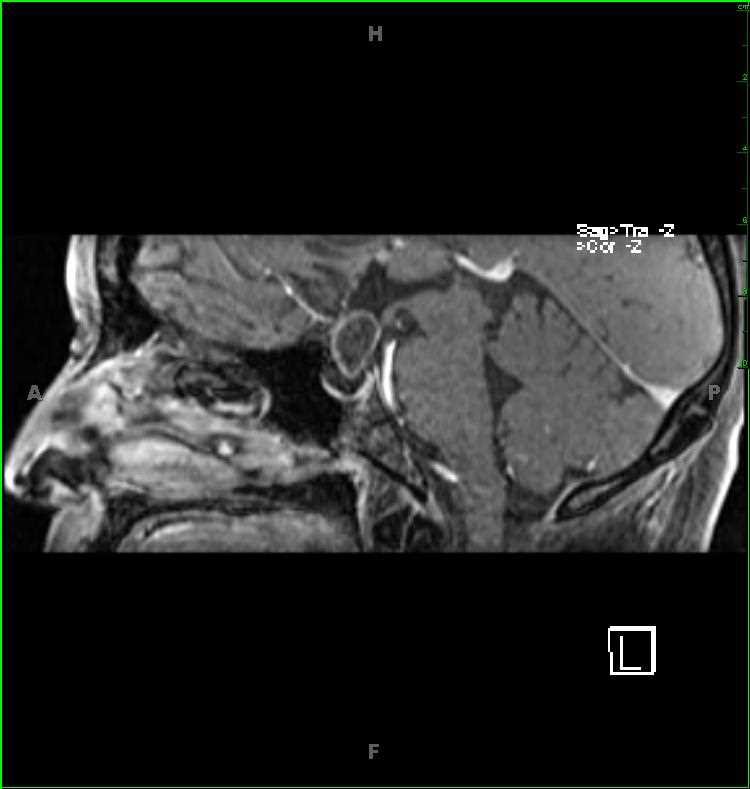
Craniopharyngioma, adamantinomatous
Note
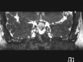
12-year-old male with chronic headaches and new onset diabetes insipidus. There is a circumscribed centrally cystic lesion arising from the anterior surface of the infindibulum. The central cystic component demonstrates signal intensity slightly greater than the surrounding CSF space on the T1-weighted image. There is FLAIR hemorrhage/proteinaceous fluid centrally. The high-resolution fluid sensitive sequences demonstrate a lesion with a thin margin abutting the posterior and inferior aspect of the optic chiasm with slight displacement and deformity. The lesion demonstrates a thin enhancing margin. The general differential includes adamantinomatous craniopharyngioma, Rathkes cleft cyst, and infindibular cyst. This is an adamantinomatous craniopharyngioma. Craniopharyngioma are though to arise via two mechanisms: as remnants of the craniopharyngeal duct, while the second is from squamous epithelial cells in the pars tuberalis of the adenohypophysis. 75% of the adamantinomatous subtype tend to be in the suprasellar compartment.
Related videos to the case

THIS IS CASE
184
OF
373