- 2
- ,
- 3
- 8
- 1
To Quiz Yourself: Select OFF by clicking the button to hide the diagnosis & additional resources under the case.
Quick Browser: Select ON by clicking the button to hide the additional resources for faster case review.
CASE NUMBER
184
Diagnosis
Craniopharyngioma, papillary
Note
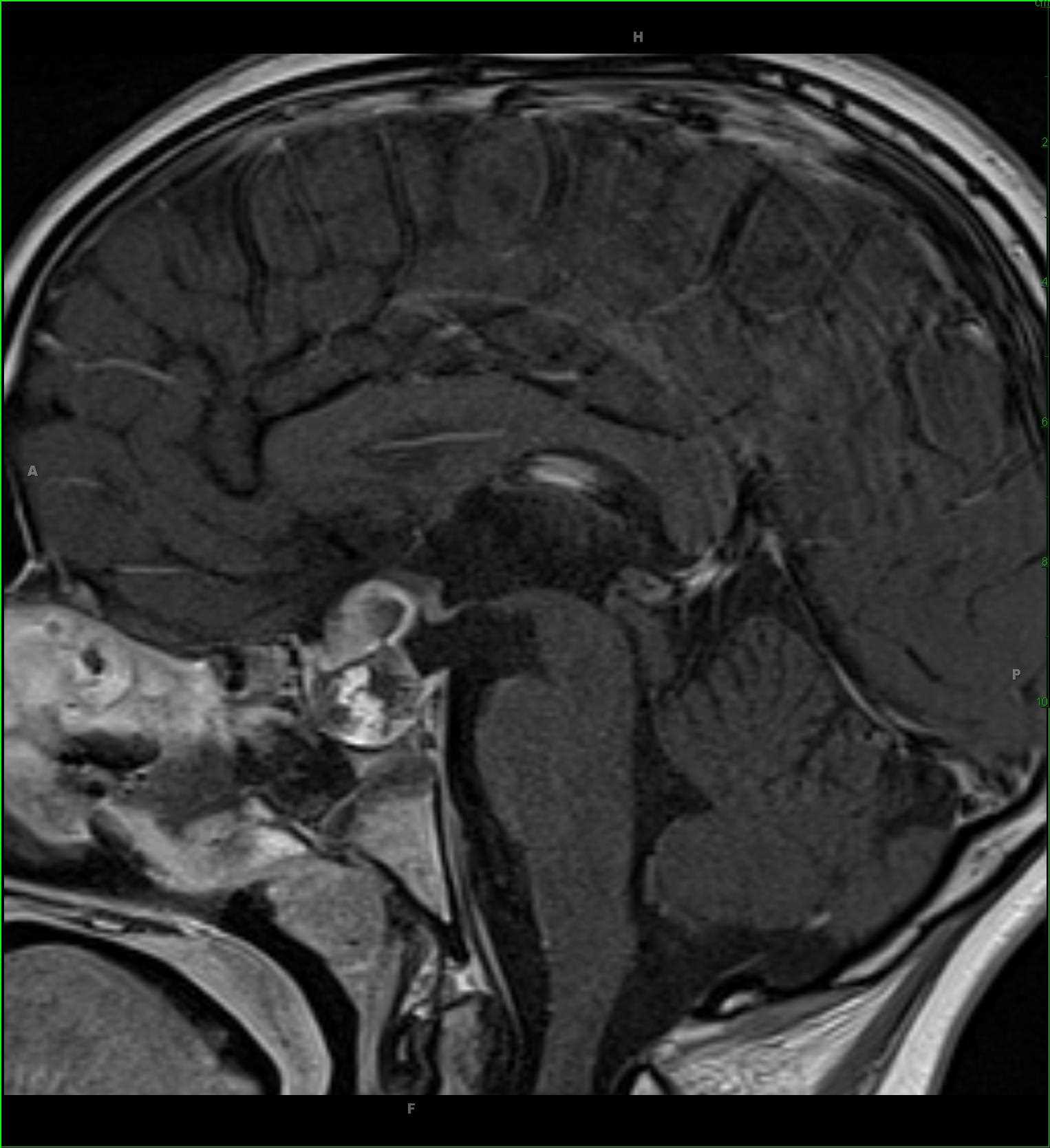
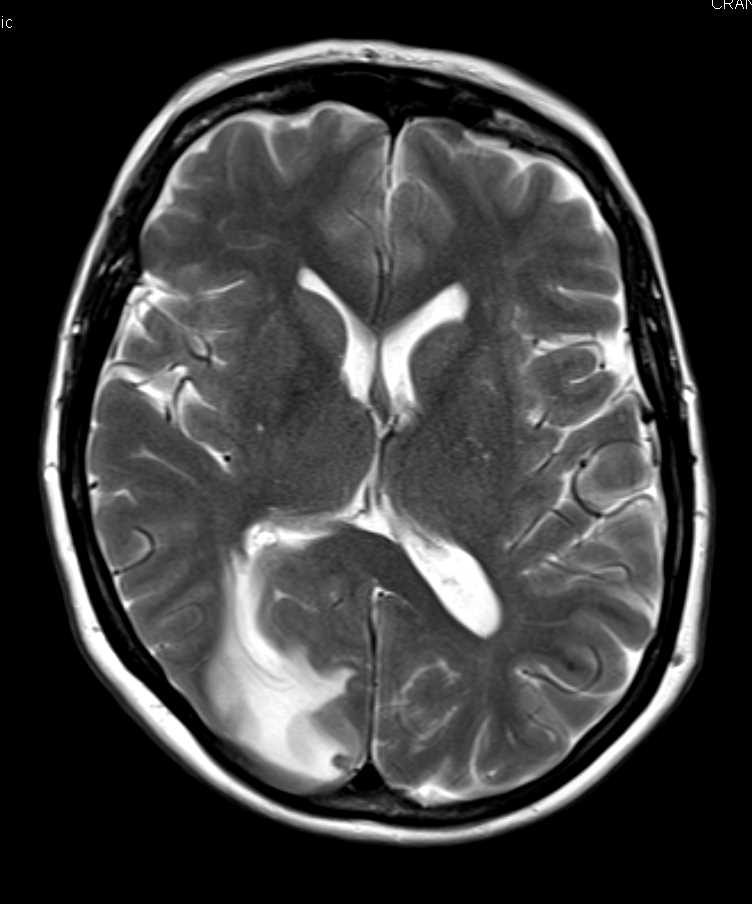
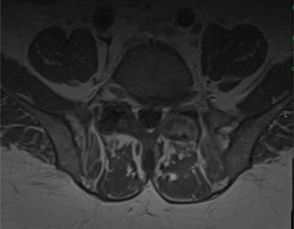
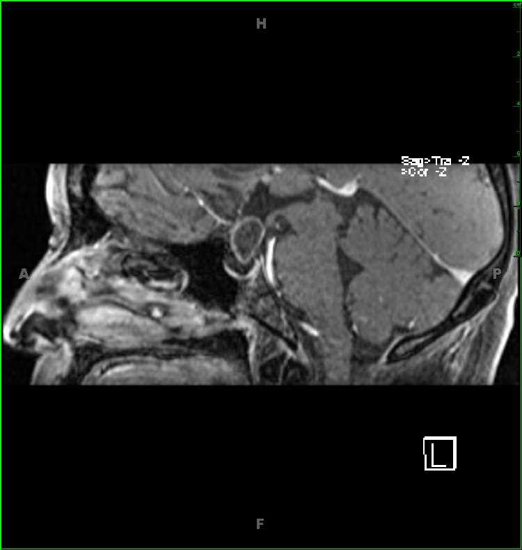
54-year-old female with chronic headaches and homonymous hemianopsia. There is a heterogeneous T1-hyper- and hypointense mass arising from the sella turcica and extending into the suprasellar compartment. Focal regions of central hemorrhage with more peripheral hemosiderin staining are identified. Nodular components of the lesion demonstrate diffusion restriction. The lesion abuts, elevates, and distorts the undersurface of the optic chiasm and prechiasmatic segments of the optic nerves superiorly. There is heterogeneous postcontrast enhancement. The findings are most compatible with a craniopharyngioma. Craniopharyngioma represent benign, partially cystic sellar/suprasellar lesions. 90% are cystic, have calcifications, and/or enhance. The adamantinomatous subtype are more common in children, with the papillary type more common in adults.
THIS IS CASE
184
OF
396


