- 2
- ,
- 2
- 4
- 6
To Quiz Yourself: Select OFF by clicking the button to hide the diagnosis & additional resources under the case.
Quick Browser: Select ON by clicking the button to hide the additional resources for faster case review.
CASE NUMBER
176


Diagnosis
Progressive Multifocal Leukoencephalopathy
Note
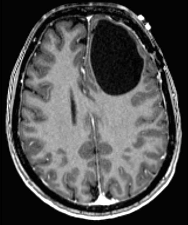
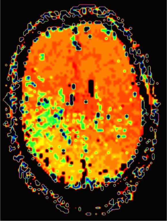
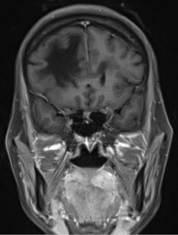
48-year-old female with a history of HIV/AIDS who presented to the ER with gradual decline in mental status. There is a confluent region of T1-hypointense and T2/FLAIR-hyperientense signal in the subcortical, periventricular and deep white matter of the right frontal lobe. There is very minimal mass effect on the overlying cerebral cortical sulci. The signal abnormality extends via the genu and anterior body of the corpus callosum to involve the white matter of the left frontal region. On the T2-weighted images, there are scattered regions of cystic change within the lesion. There are no suspicious regions of diffusion restriction or hyperperfusion to suggest high grade glioma or lymphoma. There is also no suspicious postcontrast enhancement. The imaging findings are most consistent with a diagnosis of progressive multifocal leukoencephalopathy, or PML for short. PML is a demyelinating disease resulting from oligodendrocyte infection with the JC virus. PML may be seen in any immunocompromised patient, but most commonly affects those with HIV/AIDS, transplant patients, and those with leukemia.
Related videos to the case