- 2
- ,
- 2
- 4
- 6
To Quiz Yourself: Select OFF by clicking the button to hide the diagnosis & additional resources under the case.
Quick Browser: Select ON by clicking the button to hide the additional resources for faster case review.
CASE NUMBER
173


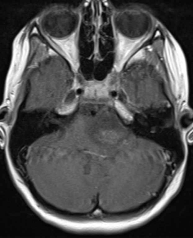

Diagnosis
Pilocytic Astrocytoma
Note
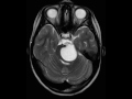
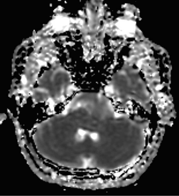
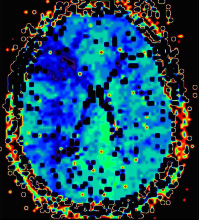
8-year-old female with worsening headaches and emesis. There is a predominantly cystic, T1-hypointense, FLAIR mildlyhyperintense, and T2-hyperintense lesion centered within the pons and left brachium pontis. The lesion extends towards the left cerebellopontine angle and prepontine space where there is partial encasement and slight rightward displacement of the basilar artery. On the postcontrast images, there is a minimally enhancing mural nodule along its posteroinferior border. The imaging findings are consistent with a pilocytic astrocytoma. Pilocytic astrocytomas are well-circumscribed, slow-growing lesions, typically demonstrating a cyst with a mural nodule. The cerebellum is the most common site of involvement, followed by the optic nerve and chiasm, and then adjacent to the third ventricle. The differential diagnosis includes medulloblastoma, ependymoma, ganglioglioma and hemangioblastoma.
Related videos to the case