- 2
- ,
- 2
- 4
- 6
To Quiz Yourself: Select OFF by clicking the button to hide the diagnosis & additional resources under the case.
Quick Browser: Select ON by clicking the button to hide the additional resources for faster case review.
CASE NUMBER
171


Diagnosis
Moyamoya Syndrome Secondary to Sickle Cell Disease
Note
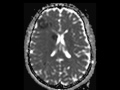
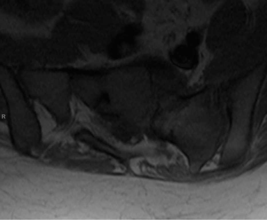
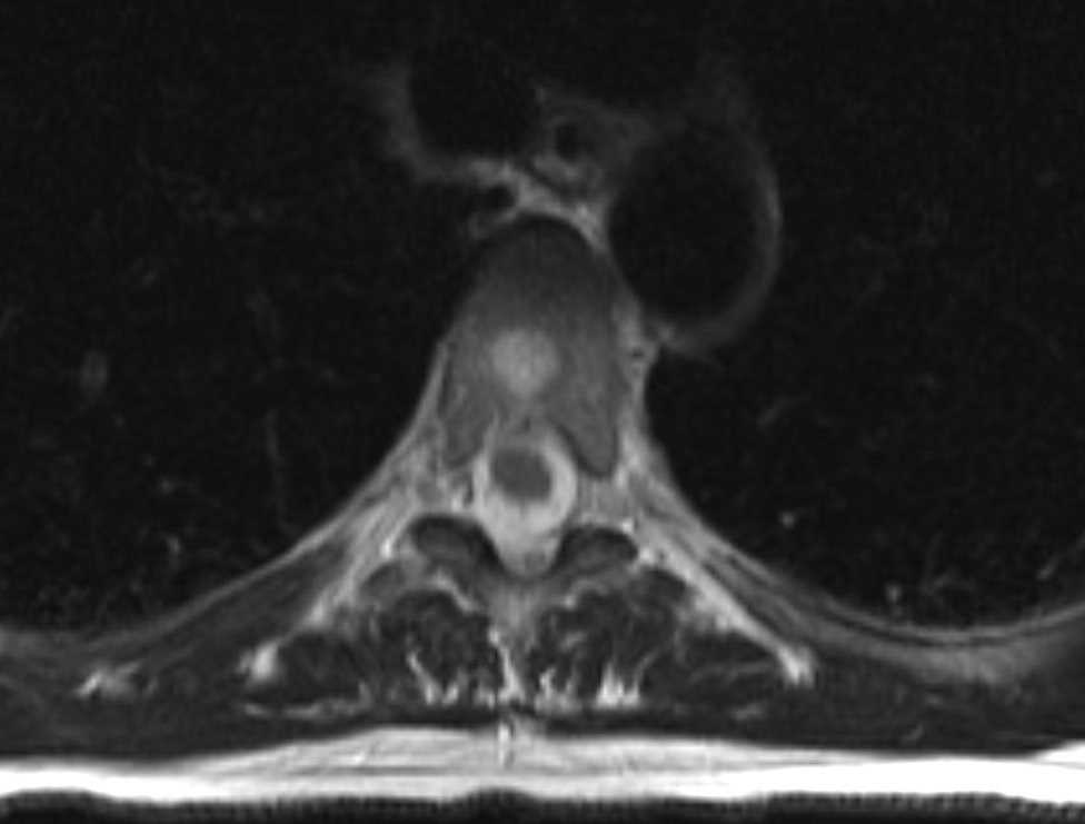
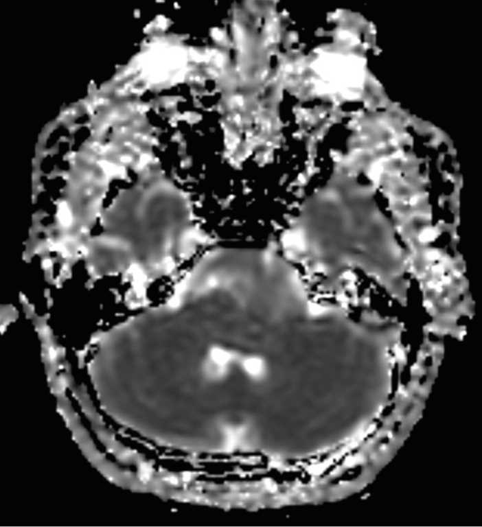
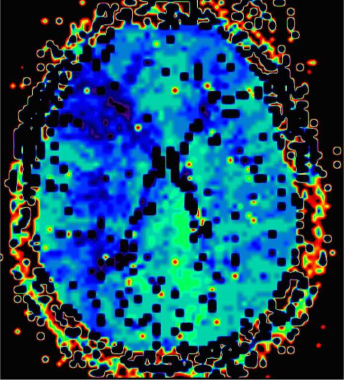
13-year-old female with history of sickle cell disease and prior left middle cerebral artery infarct presents for new altered mental status. There are numerous punctate and more confluent acute infarcts within the right caudate head and the watershed territory of the right MCA and ACA territories. The arterial spin label perfusion maps demonstrate large regions of ischemic penumbra emanating from the watershed zones between the right ACA and MCA as well as the right PCA and MCA territories. The time to peak contrast enhanced perfusion maps also demonstrate an ischemic penumbra in the watershed zone between the left ACA and MCA. The 3D MIP image from the 3D time of flight MRA demonstrates numerous tiny collateral vessels extending from the supraclinoid termini through the first segments of the right and left middle cerebral arteries with additional involvement of the first segment of the right posterior cerebral artery. The findings are compatible with moyamoya syndrome in the setting of sickle cell disease. Myoamoya disease describes the vascular phenomenon in the idiopathic or familial setting. The syndrome can be seen secondary to radiation, infection, connective tissue disorders, fibromuscular dysplasia, Down syndrome and atherosclerosis among many other causes.
Related videos to the case