- 2
- ,
- 2
- 4
- 6
To Quiz Yourself: Select OFF by clicking the button to hide the diagnosis & additional resources under the case.
Quick Browser: Select ON by clicking the button to hide the additional resources for faster case review.
CASE NUMBER
167


Diagnosis
Laminar Necrosis
Note
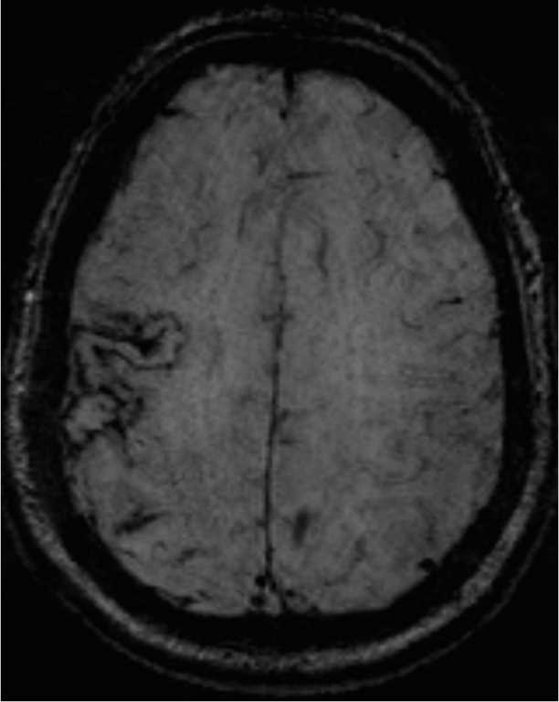
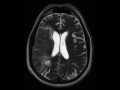
64-year-old male with a history of a large right middle cerebral artery infarct. The fluid sensitive images demonstrate residual edema and loss of gray-white matter differentiation within the right MCA territory. There is also residual effacement of the overlying cerebral cortical sulci with very slight effacement of the body and atrium of the right lateral ventricle. The susceptibility weighted images demonstrate curvilinear regions of signal loss along the cortical gray matter of the right frontoparietotemporal regions as well as of the insula. Findings are compatible with cortical laminar necrosis in the setting of recent infarct. Cortical laminar necrosis is secondary to selective vulnerability or watershed cortical layers. Neurons and glial cells are equally affected. Causes include hypoperfusion, hypoxia, status epileptics, hypoglycemia and severe anemia.
Related videos to the case

THIS IS CASE
167
OF
373