- 2
- ,
- 2
- 4
- 6
To Quiz Yourself: Select OFF by clicking the button to hide the diagnosis & additional resources under the case.
Quick Browser: Select ON by clicking the button to hide the additional resources for faster case review.
CASE NUMBER
165


Diagnosis
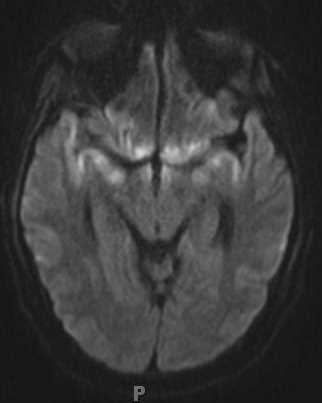
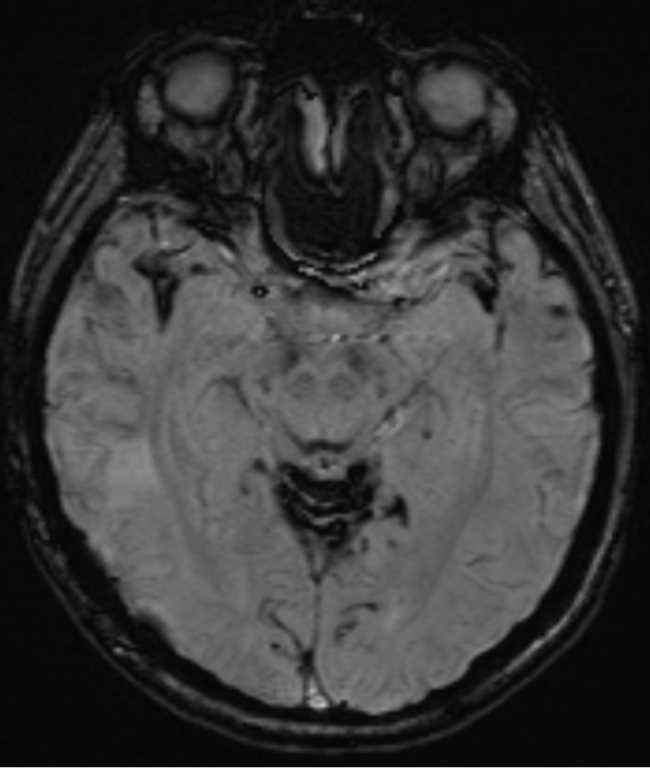
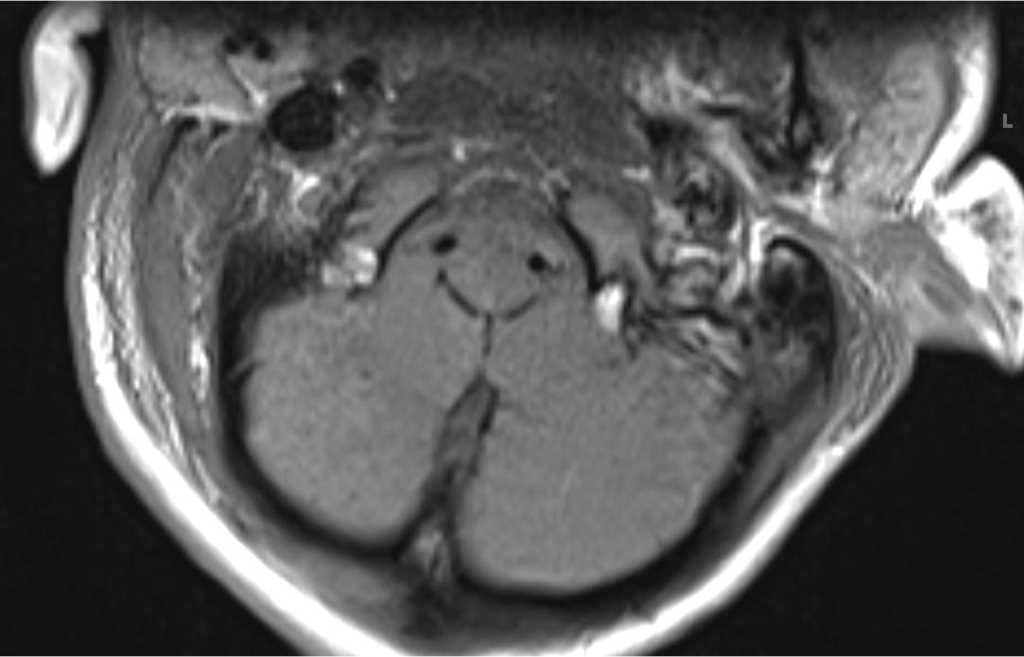
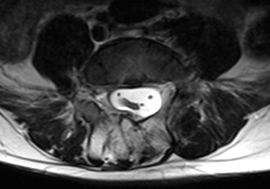
Hydromyelia and Diplomyelia
Note
53-year-old female with a history of hydroomyelia and diplomyelia. The axial T2-weighted images demonstrate a well defined T2-hyperintense cystic cavity within the central cord substance of the lower thoracic and upper lumbar spinal regions compatible with a dilated central canal. The focal region of hydromyelia is associated with cord expansion and thinning of the overlying cord substance. More inferiorly to this level, there is division of the cord into two hemicords by an osseous septum. On myelography, there were two ventral and dorsal nerve roots from each hemicord, compatible with a diagnosis of diplomyelia. The axial T1-weighted image demonstrates fatty marrow extending into the osseous septum. Hydromyelia results from cystic dilatation of the central canal, while syringomyelia is a cystic or multicystic spinal cord cavity not contiguous with the central canal. The differential diagnosis for a cystic lesion of the lower thoracic and upper lumbar spinal cord includes ventriculus terminalis, cystic spinal cord tumor, and myelomalacia.
Related videos to the case


THIS IS CASE
165
OF
373