- 2
- ,
- 2
- 4
- 6
To Quiz Yourself: Select OFF by clicking the button to hide the diagnosis & additional resources under the case.
Quick Browser: Select ON by clicking the button to hide the additional resources for faster case review.
CASE NUMBER
161


Diagnosis
Adamantinomatous Craniopharyngioma
Note
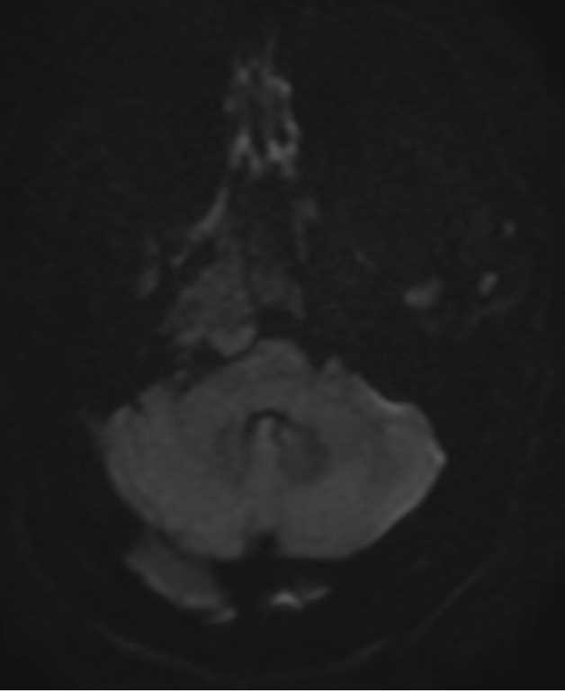
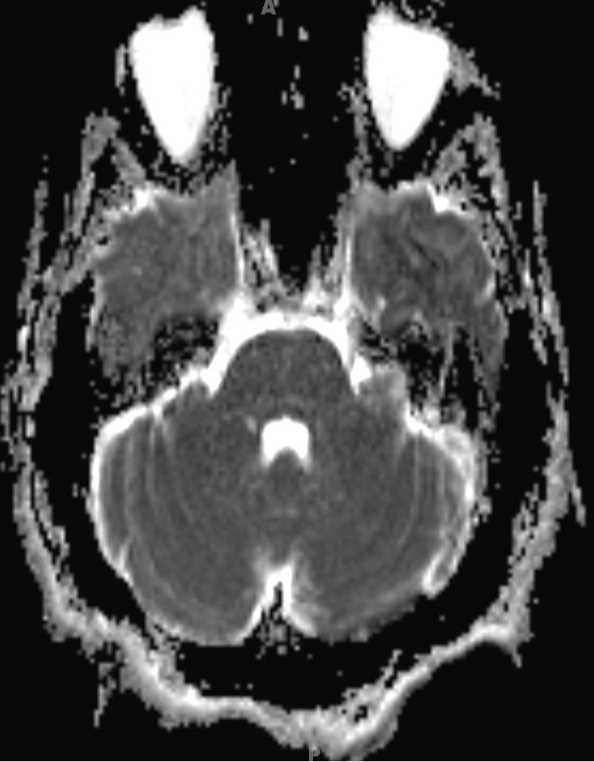
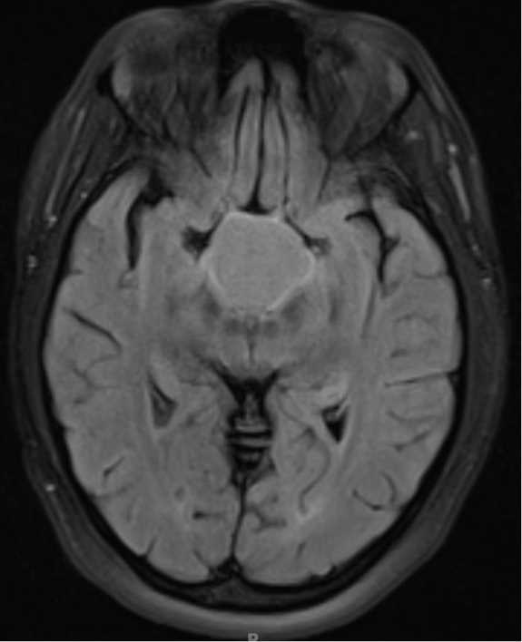
16-year-old male with bitemporal hemianopsia and chronic headaches. There is circumscribed T1-hypointense, T2-hyperintense mass centered within the sellar/suprasellar region. The lesion demonstrates T2/FLAIR-hyperintense signal, facilitated diffusion, and a thin peripheral rim of postcontrast enhancement. There are lobular cystic components which extend into the enlarged sella turcica. This is a case of an adamantinomatous craniopharyngioma. Craniopharyngiomas occur with a bimodal distribution. The first peak occurs between 10 and 14 years of age while the second occurs in middle-aged adutls. Pediatric cases are mostly composed of the adamantinomatous subtype while the papillary subtype comprises the majority of cases in middle-aged adults. Cases occur in males and females about equally. Differential includes Rathke cleft cyst, cystic pituitary adenoma, epidermoid and intracranial teratoma.
Related videos to the case