- 2
- ,
- 2
- 4
- 6
To Quiz Yourself: Select OFF by clicking the button to hide the diagnosis & additional resources under the case.
Quick Browser: Select ON by clicking the button to hide the additional resources for faster case review.
CASE NUMBER
159


Diagnosis
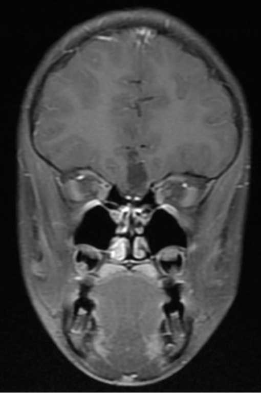
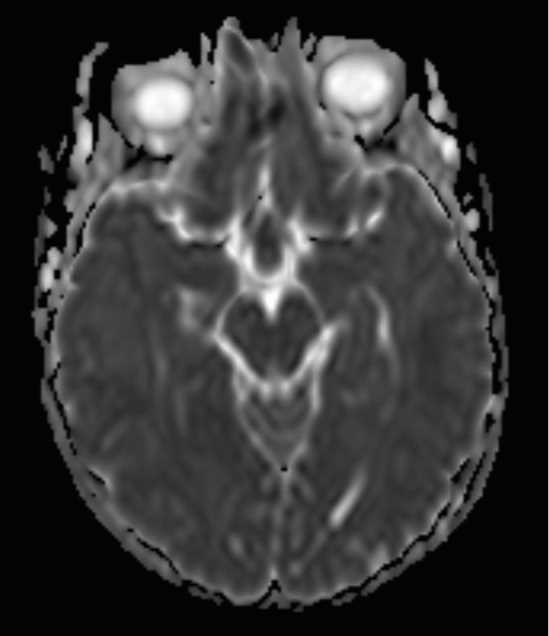
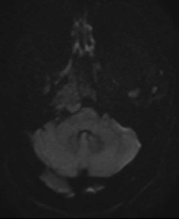
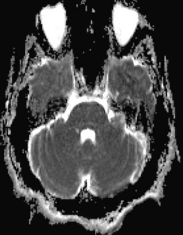
Cerebellopontine Angle Dermoid
Note
This is a case of a left cerebellopontine angle dermoid. There is a nonenhancing T1-isointense lesion with lobular margins in the left cerebellopontine angle. The lesion is T2-hyperintense with scattered cystic components. It extends to the posterior margin of the porus acoustics and minimally extends into the left internal auditory canal. The lesion is FLAIR-hyperintense and mildly restrict diffusion. The dermoid represents an inclusion of ecteodermal tissues within the CNS. Dermoids contain squamous epithelium and associated dermal appendages. Dermoids are typically hyperintense on T1, heterogeneous on T2, and with a striated or layered appearance. The differential diagnosis includes craniopharyngioma, arachnoid cyst, and lipoma. Dermoids are usually well circumscribed while epidermoids are more amorphous.
Related videos to the case