- 2
- ,
- 2
- 4
- 6
To Quiz Yourself: Select OFF by clicking the button to hide the diagnosis & additional resources under the case.
Quick Browser: Select ON by clicking the button to hide the additional resources for faster case review.
CASE NUMBER
158


Diagnosis
Planum Sphenoidale Dermoid
Note
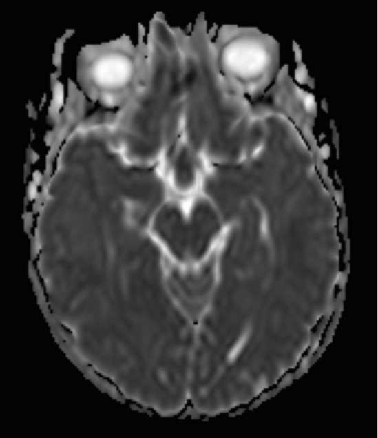
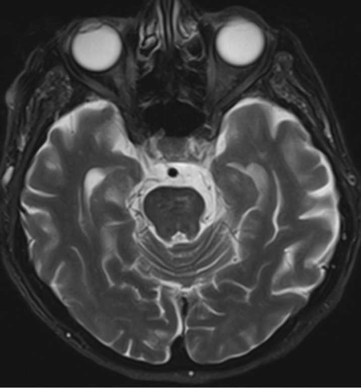
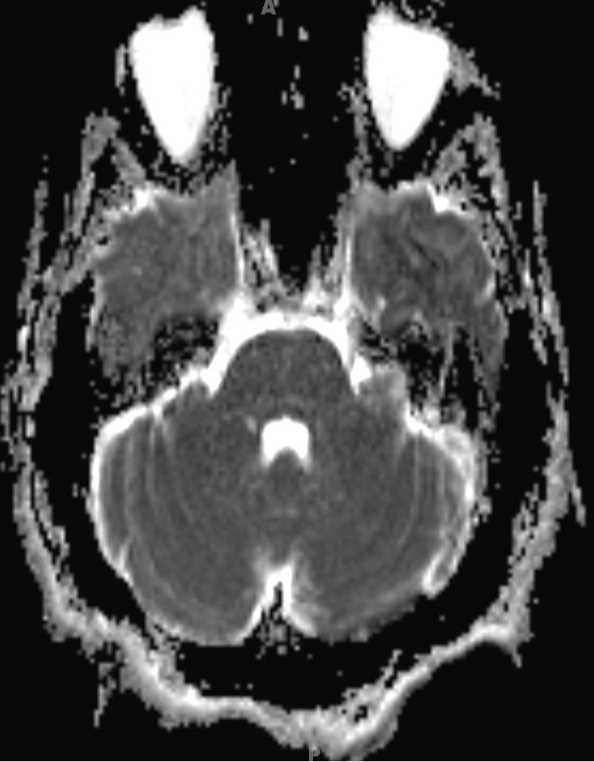
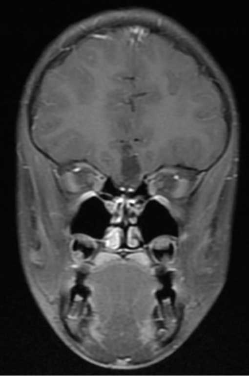
Centered within the anterior interhemispheric fissure and arising directly superior to the planum sphenoidale, there is an ovoid T1 predominantly isointense lesion. There are a few scattered regions of T1-hyperintense signal within the lesion. The heterogenous nature of the lesion and origin from the planum sphenoidal are better demonstrated on the sagittal T1-weighted image. There is hypointense signal on the FLAIR-weighted image, and hyperintense signal on the T2-weighted images. On the fat suppressed, post contrast T1-weighted image, there is diffuse signal loss. The findings are compatible with a planum sphenoidale dermoid. The dermoid represents an inclusion of ectodermal tissues within the CNS. Dermoids contain squamous epithelium and associated dermal appendages. Dermoids are typically hyperintense on T1, heterogeneous on T2, and with a striated or layered appearance. The differential diagnosis includes craniopharyngioma, arachnoid cyst, and lipoma.
Related videos to the case


THIS IS CASE
158
OF
373