- 2
- ,
- 2
- 4
- 6
To Quiz Yourself: Select OFF by clicking the button to hide the diagnosis & additional resources under the case.
Quick Browser: Select ON by clicking the button to hide the additional resources for faster case review.
CASE NUMBER
156


Diagnosis
Bilateral ICA Occlusion
Note
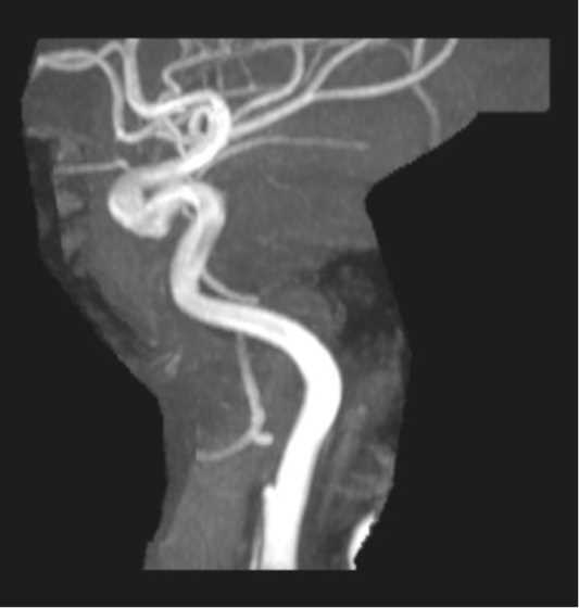
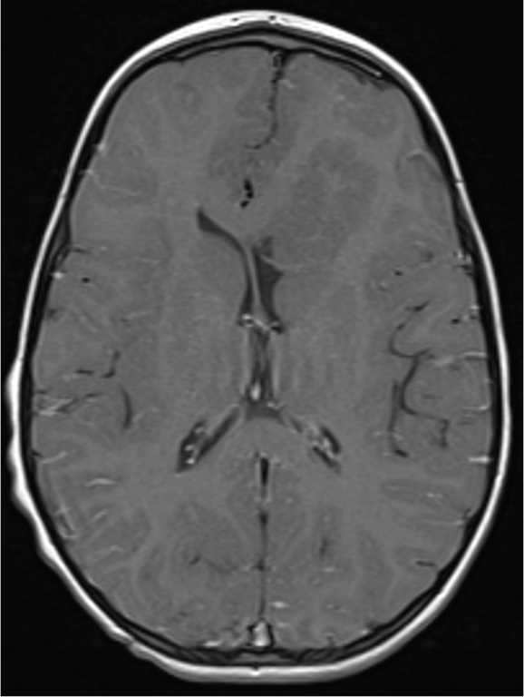
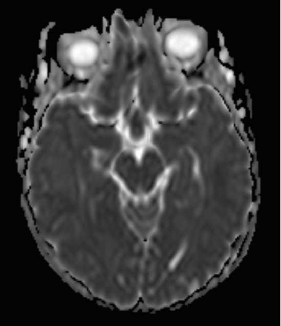
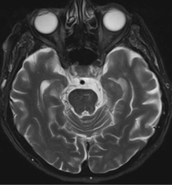
This 78-year-old female with history of hypertension, diabetes, and smoking presented for new onset altered mental status. The first three axial T2-weighted images demonstrate absence of flow voids within the right and left ICA petrous to paraclinoid segments. The 3-D time of flight MRA image demonstrates flow related signal in the distal basilar artery. No flow related signal is identified in either the right or left ICA cavernous segment. The findings are summarized on the 3-D MIP reconstruction where neither internal carotid segment is identified. There is abrupt cutoff of the left distal common carotid artery. Bilateral ICA occlusion typically presents in patients with a history of smoking and diabetes.
Related videos to the case