- 2
- ,
- 2
- 4
- 6
To Quiz Yourself: Select OFF by clicking the button to hide the diagnosis & additional resources under the case.
Quick Browser: Select ON by clicking the button to hide the additional resources for faster case review.
CASE NUMBER
145


Diagnosis
Chronic Lymphocytic Leukemia of the Maxillofacial Region
Note
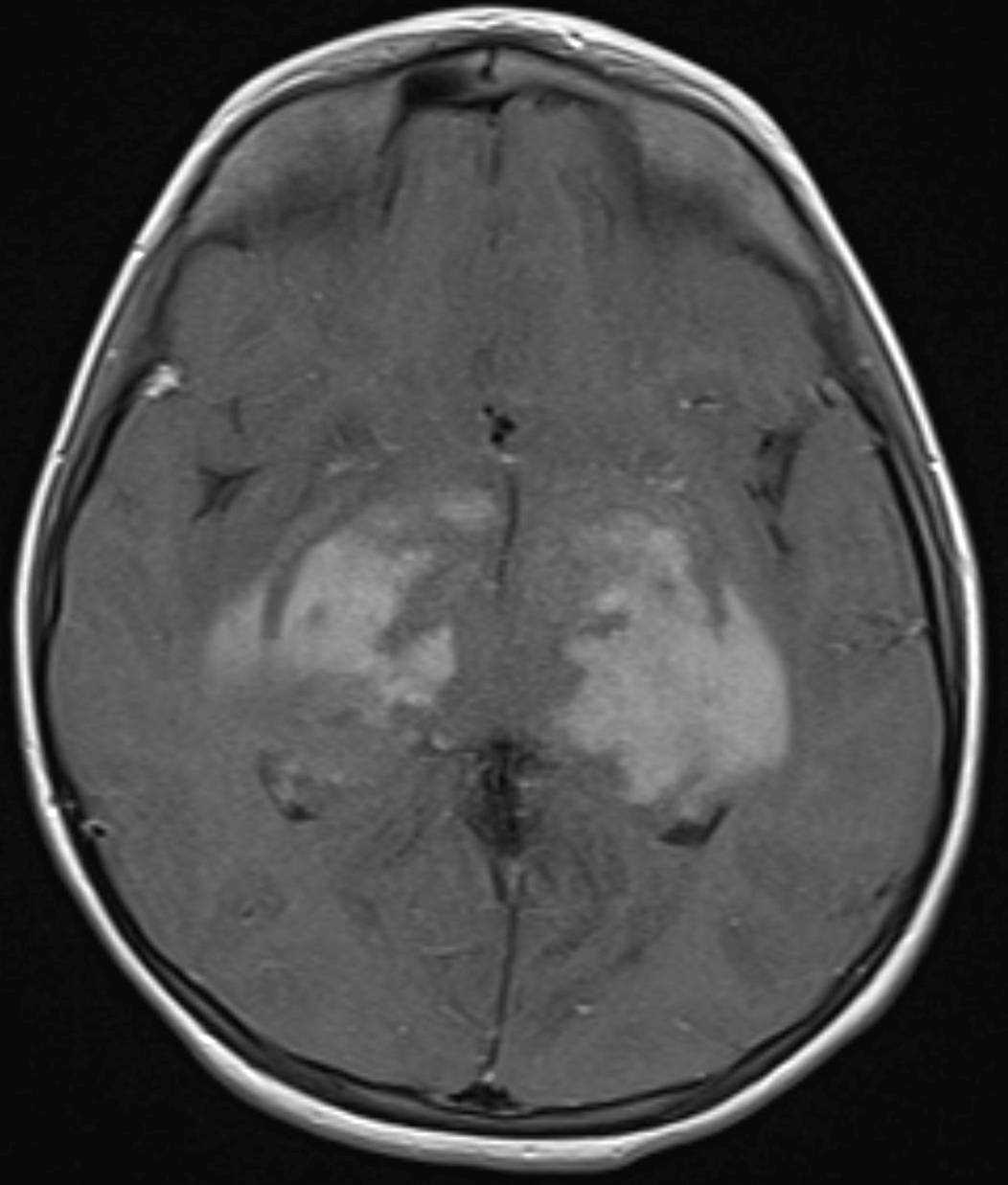
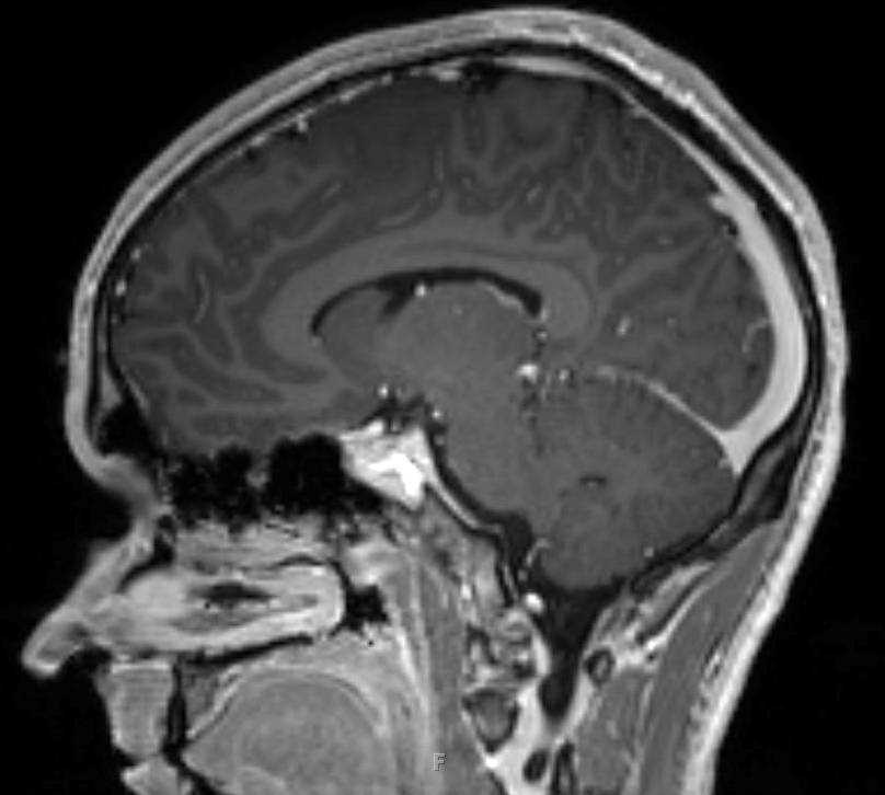
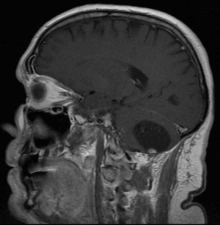
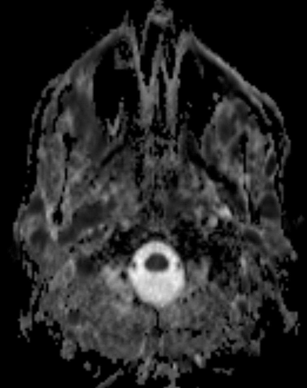
78-year-old male who presented for chronic headaches and fatigue. Numerous T2/FLAIR minimally hyperintense, rounded, soft tissue lesions are identified involving the bilateral temporal fossa, masticator, parapharyngeal and parotid spaces. There are infiltrating T2/FLAIR hyperintense lesions involving the left medial rectus muscle, extraconal fat, pterygoid muscle complexes and left adenoid. The lesions are more apparent on the diffusion weighted images where there is hyperintense signal and on the ADC maps where there are low values. This was a case of biopsy proven chronic lymphocytic leukemia of the maxillofacial region. Chronic lymphocytic leukemia is the most common type of leukemia to affect elderly adults.
Related videos to the case