- 2
- ,
- 2
- 4
- 6
To Quiz Yourself: Select OFF by clicking the button to hide the diagnosis & additional resources under the case.
Quick Browser: Select ON by clicking the button to hide the additional resources for faster case review.
CASE NUMBER
144


Diagnosis
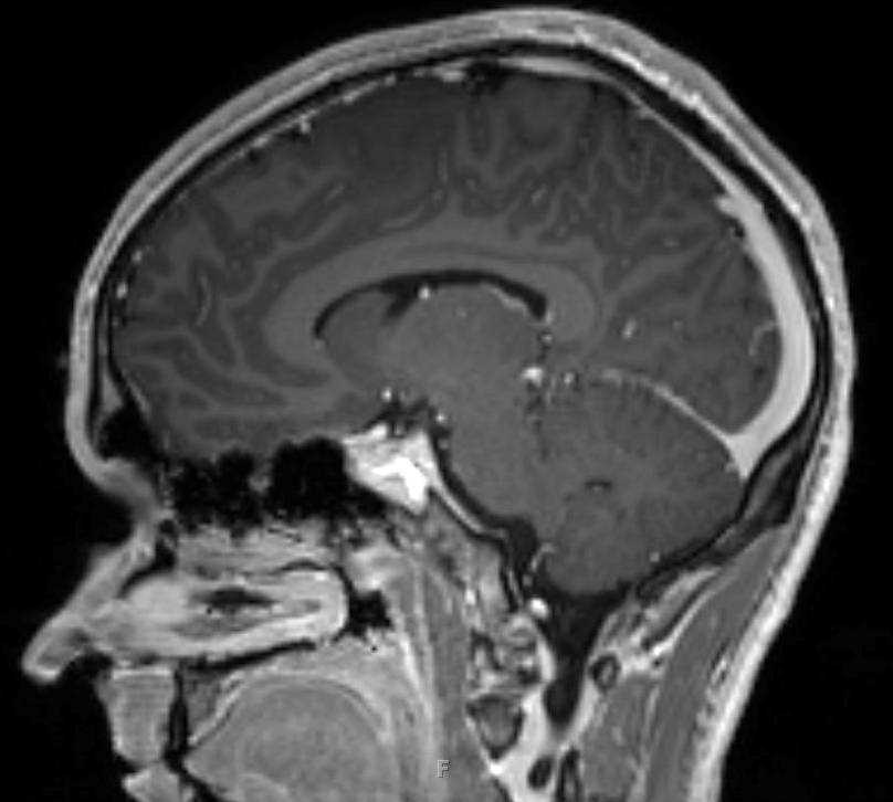
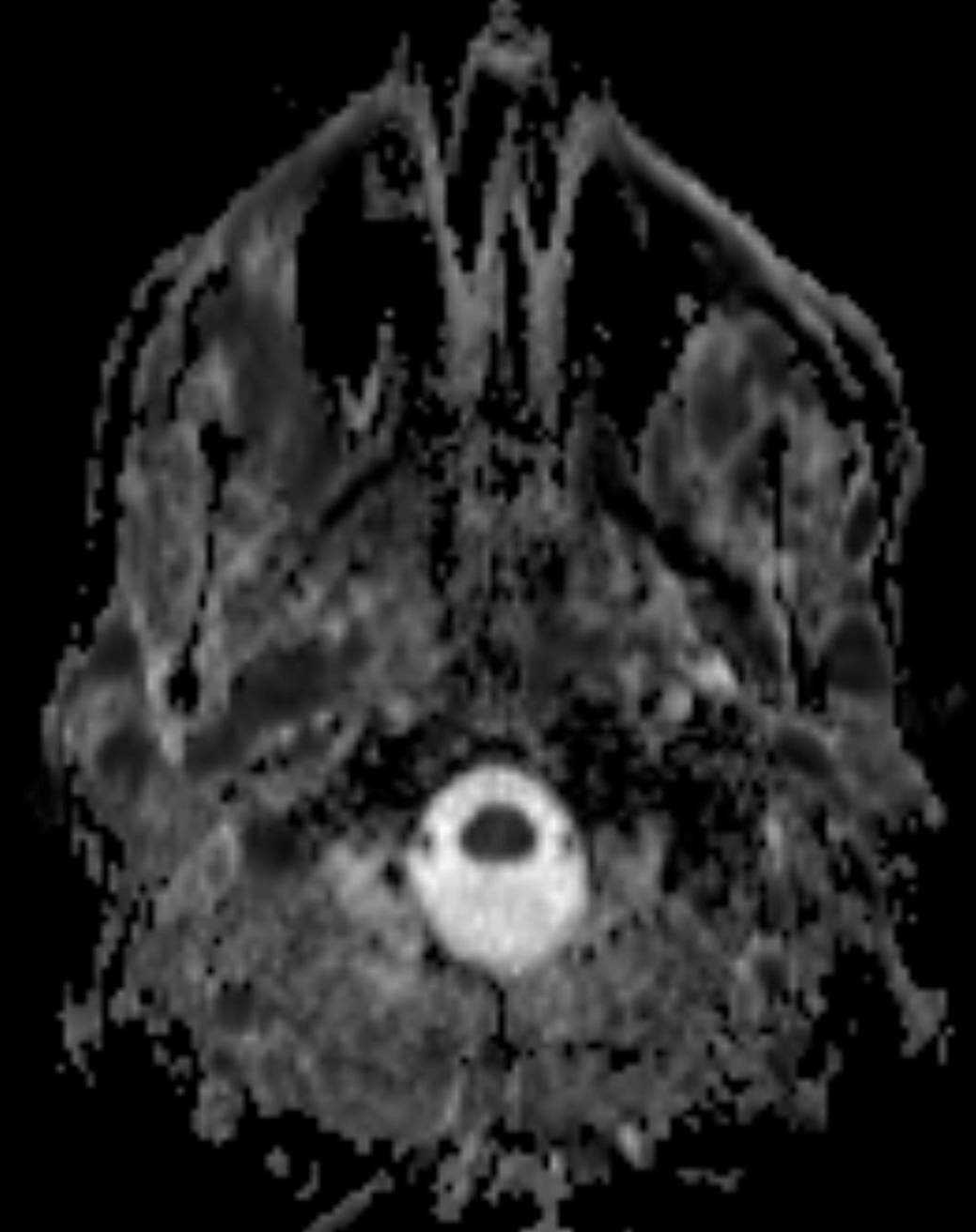
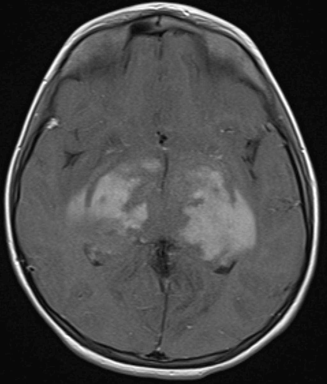
Optic Pathway Glioma
Note
12-year-old male presented with a long standing history of progressive loss of vision. The prechiasmatic segments of the optic nerves are thickened and hyperintense on the T2-weighted images. There is also thickening and T2 hyperintense signal within the optic chasm which contacts and elevates the A1 segment of the right and left anterior cerebral arteries. More posteriorly, there is a diffuse infiltrating mass which invades and expands the bilateral thalami, optic radiations, basal ganglia, medial temporal lobes and hippocampi. The findings result in diffuse effacement of the suprasellar and ambient cisterns with mild mass effect and edema within the midbrain. There are globular regions of avid enhancement on the post contrast images within the thalami, basal ganglia, and optic radiations within the temporal lobes. The imaging findings are compatible with an optic pathway glioma. Typical imaging features include fusiform enlargement of the optic nerve with variable involvement of the more posterior optic pathways. The lesions are variably hyperintense on T2 weighted images, and enhancement ranges from minimal to intense.
Related videos to the case