- 2
- ,
- 2
- 4
- 6
To Quiz Yourself: Select OFF by clicking the button to hide the diagnosis & additional resources under the case.
Quick Browser: Select ON by clicking the button to hide the additional resources for faster case review.
CASE NUMBER
133


Diagnosis
Vestibular Schwannoma
Note
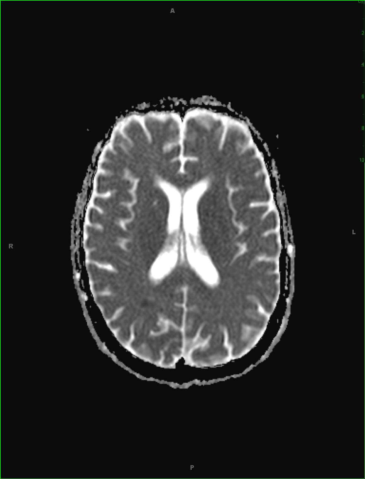
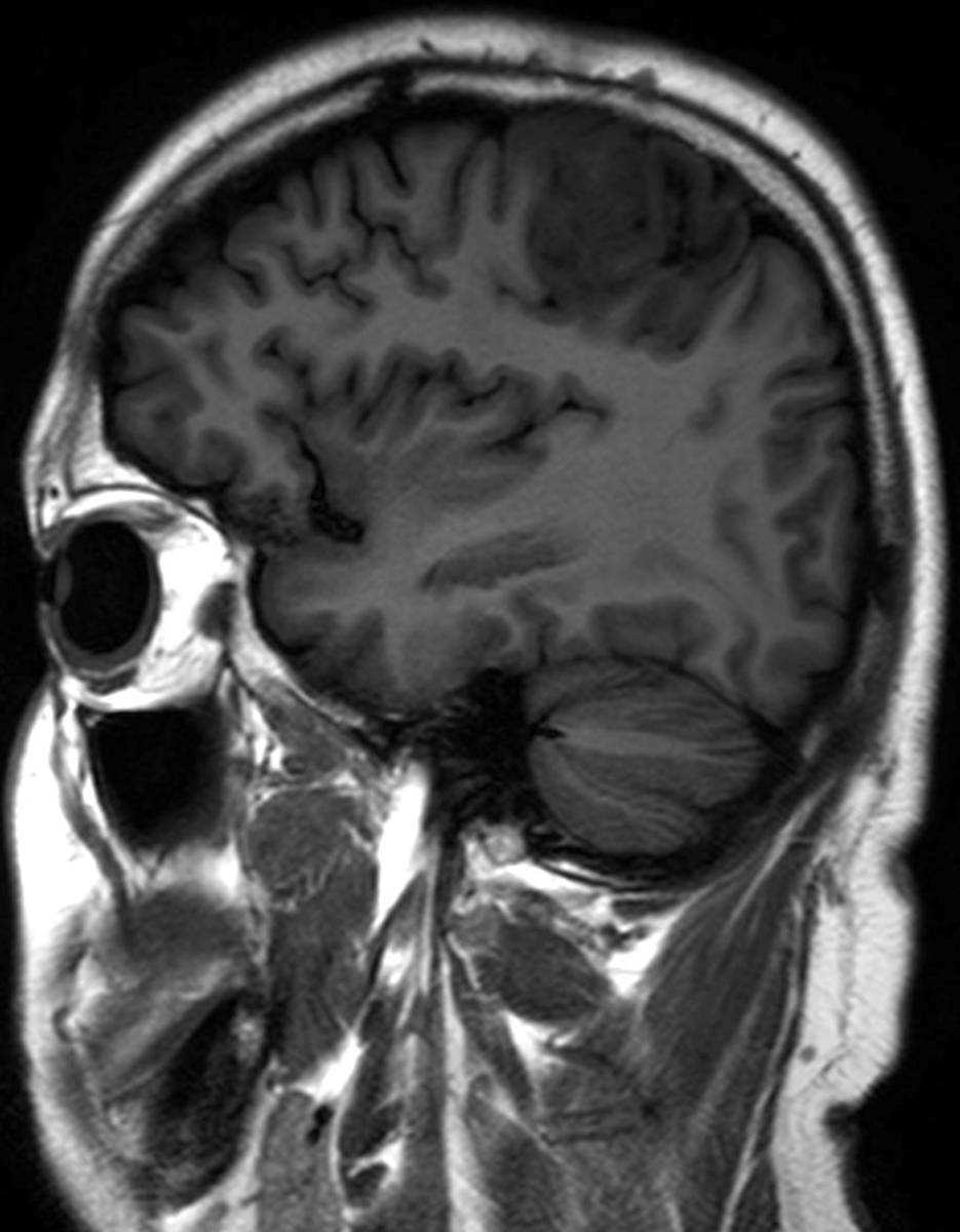
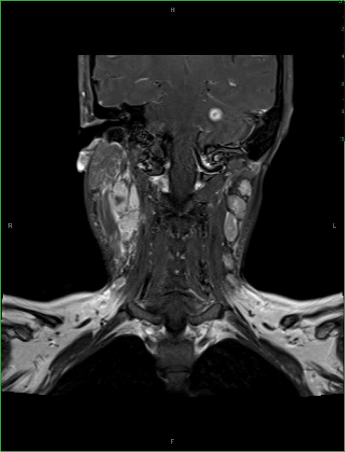
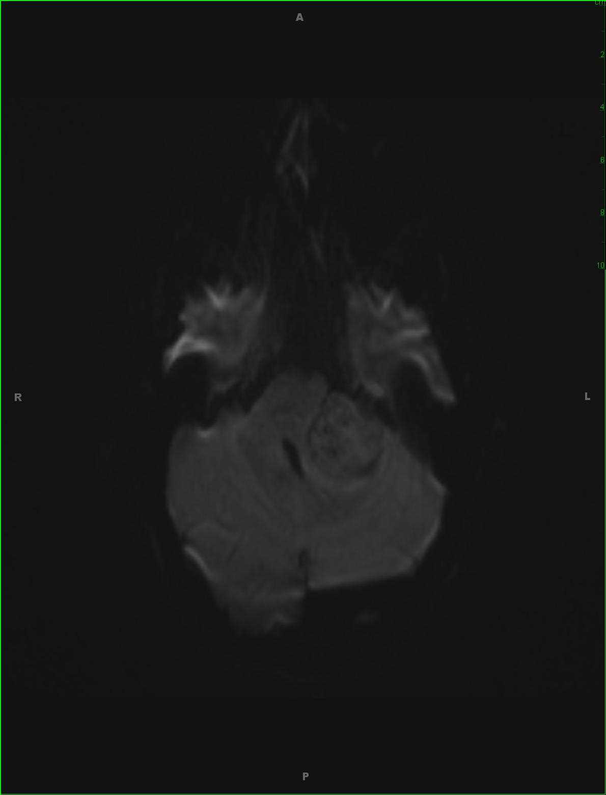
This is a case of a vestibular schwannoma in a 28-year-old female with left-sided tinnitus and hearing loss. The first image, a sagittal T1-weighted sequence off midline, demonstrates a heterogeneous T1-hypointense mass filling the left cerebellopontine angle. The second and third images, axial FLAIR and T2-weighted sequences, demonstrates heterogeneous, predominantly hyperintense signal within the mass arising in the left internal auditory canal and filling the left cerebellopontine angle. The hyperintense components of the lesion likely reflect the presence of Antoni B, myxomatous, tissue at those sites. There are no suspicious findings on the diffusion weighted images, four and five. The postcontrast fat-saturated T1-weighted sequence, image 6, demonstrates an avidly enhancing mass filling and expanding the left internal auditory canal and porus acoustics.
Related videos to the case