- 2
- ,
- 2
- 4
- 6
To Quiz Yourself: Select OFF by clicking the button to hide the diagnosis & additional resources under the case.
Quick Browser: Select ON by clicking the button to hide the additional resources for faster case review.
CASE NUMBER
131


Diagnosis
Low-grade Oligoastrocytoma
Note
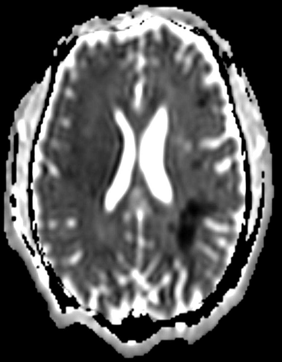
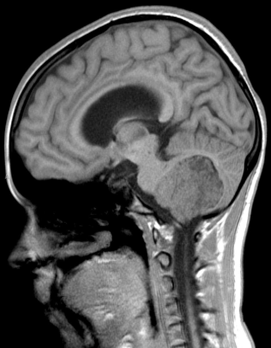
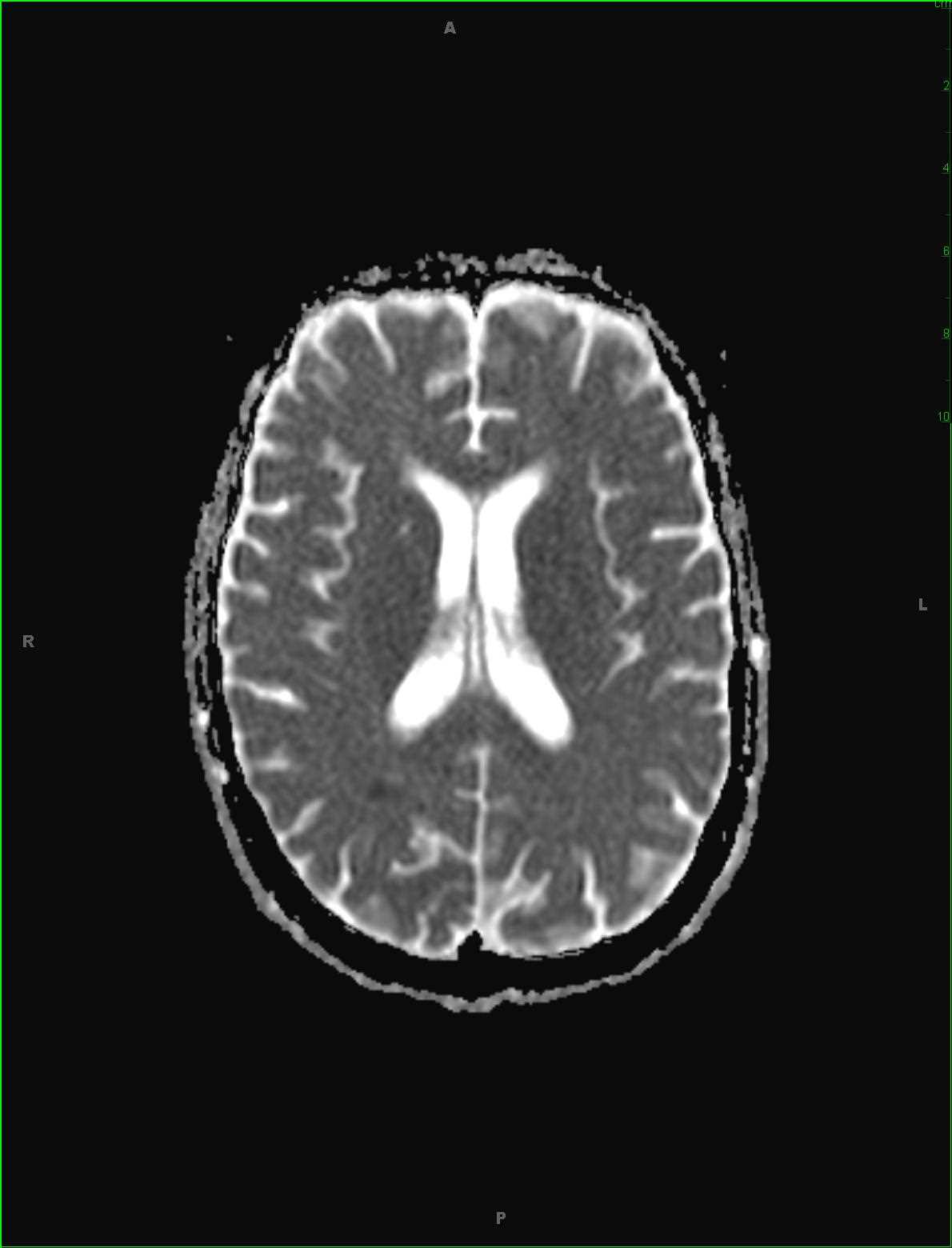
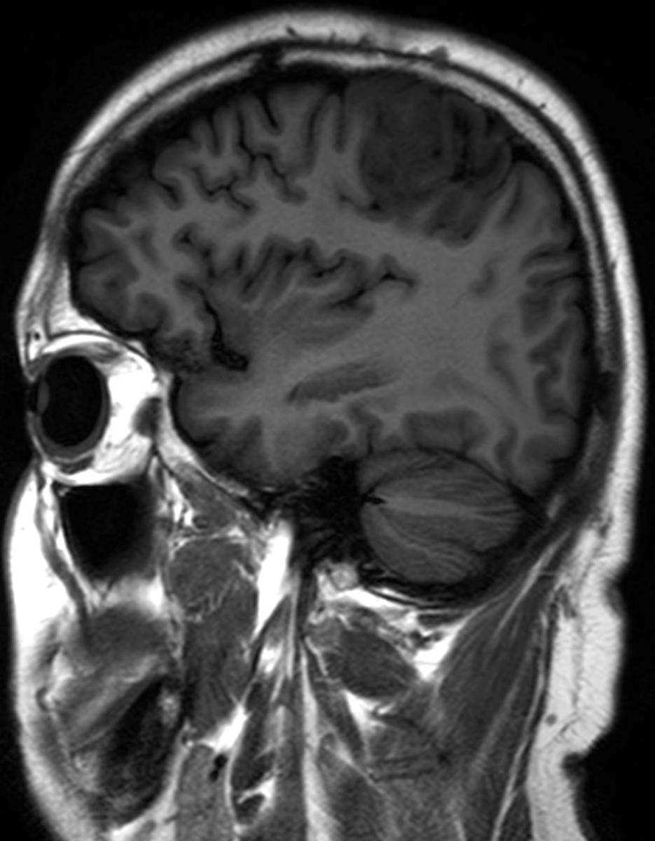
This is a case of a 34-year-old male presenting with new right-sided tonic clonic seizures resulting from an left-sided oligoastrocytoma centered in the left prefrontal gyrus. The lesion involves both the cortical grey matter and subcortical white matter with associated gyral expansion on the sagittal T1-weighted sequence, image 1. There is increased FLAIR signal intensity at the lesion site, image 2, which reflects a combination of vasogenic edema and infiltrating tumor. There are a few focal regions of signal loss along the cortical sulci, which reflects minimal mineralization and/or calcification. The DWI and ADC maps, images 3 and 4, demonstrate no diffusion restriction. The axial T2-weighted sequence, image 5, redemonstrates the hyperintense signal previously demonstrated on the FLAIR image. There is subtle effacement of the overlying cortical sulci secondary to the gyral swelling. The final image is a post contrast T1-weighted sequence, demonstrating avid but heterogeneous postcontrast enhancement at the anterior aspect of the lesion. Oligoastrocytomas demonstrate a mixed cellular histology, usually acting as medium-grade neoplasms with preferential localization to the frontal and temporal regions. There is a high rate of local recurrence. It is more common to find postcontrast enhancement as compared to calcification or mineralization in these tumors.
Related videos to the case