- 2
- ,
- 2
- 4
- 6
To Quiz Yourself: Select OFF by clicking the button to hide the diagnosis & additional resources under the case.
Quick Browser: Select ON by clicking the button to hide the additional resources for faster case review.
CASE NUMBER
125


Diagnosis
Embolic Infarcts
Note
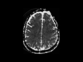
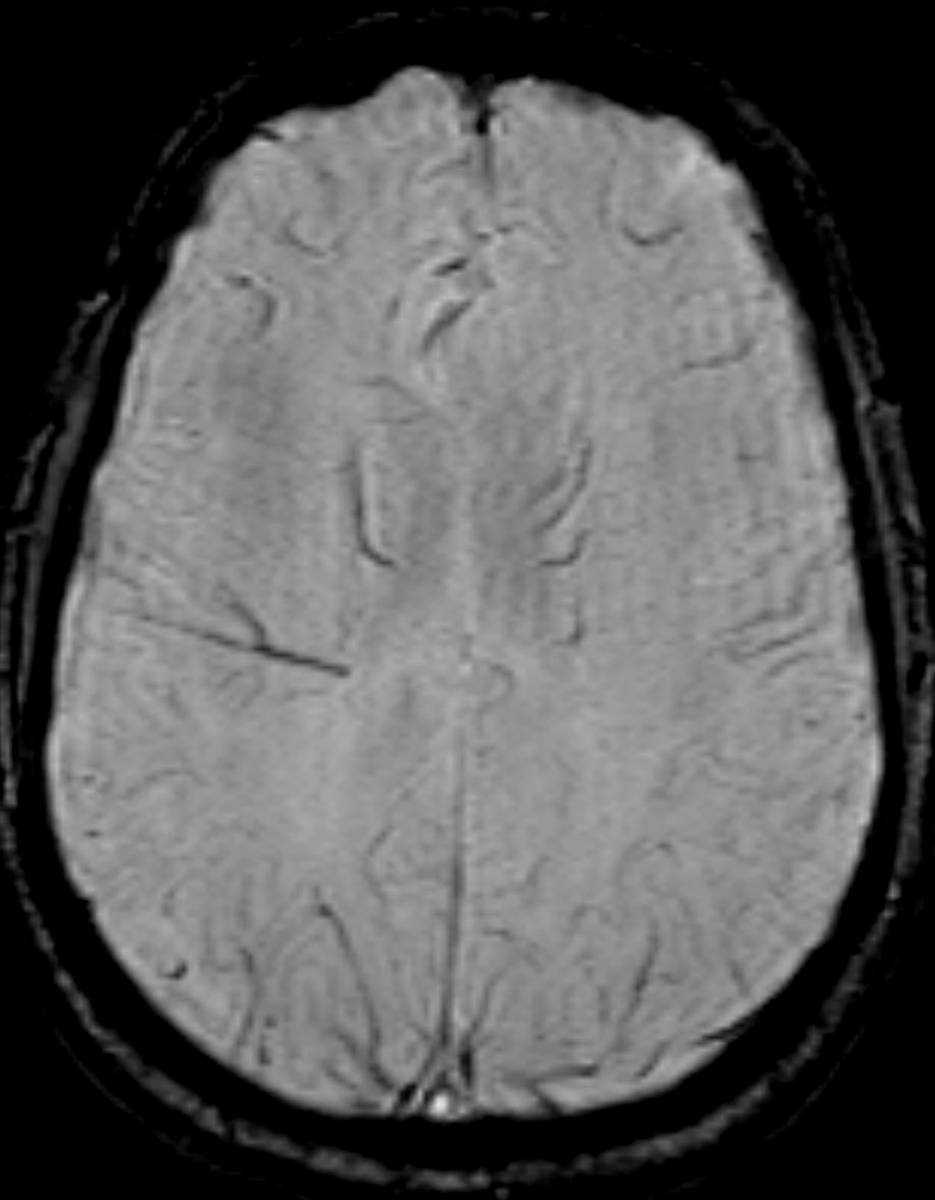
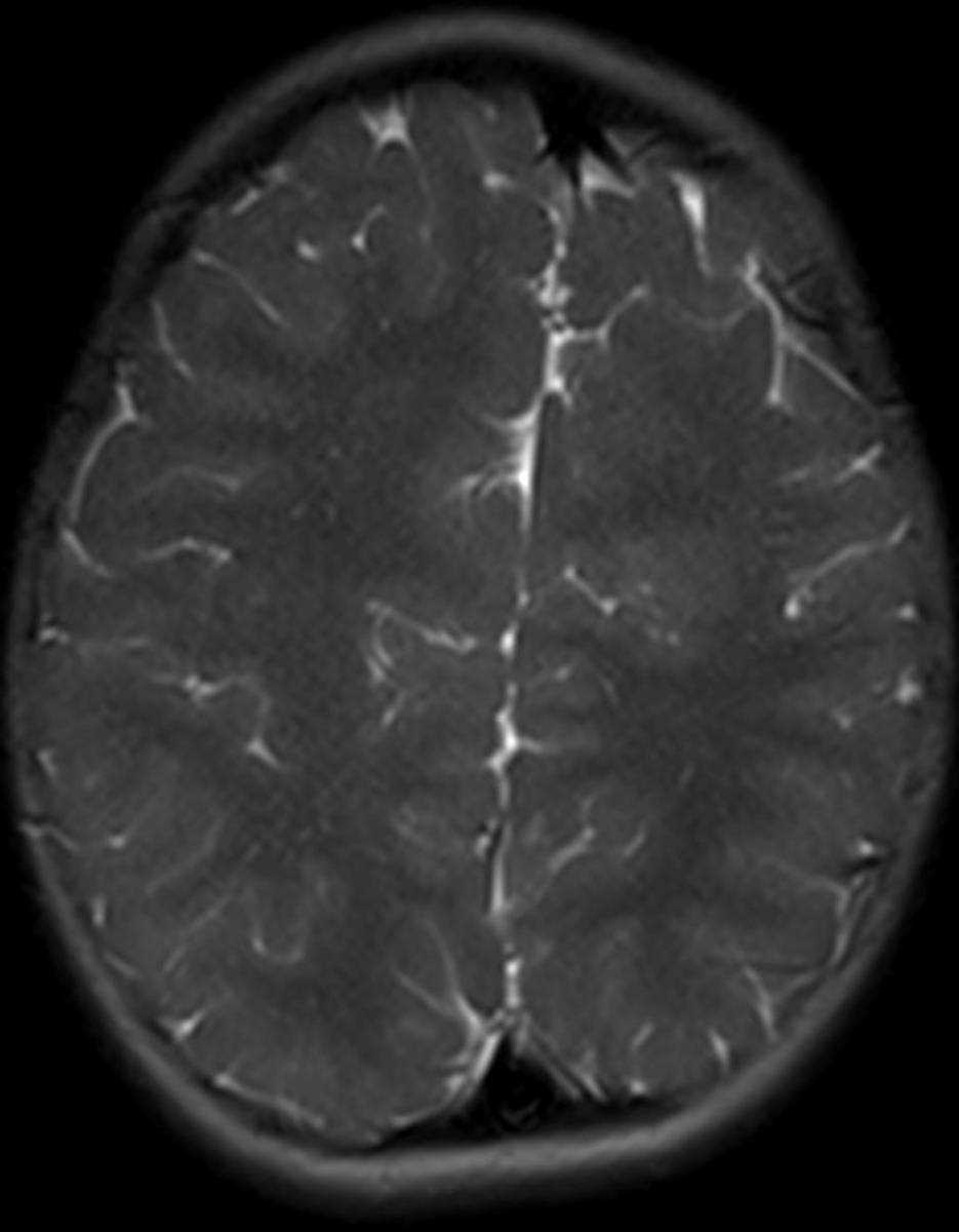
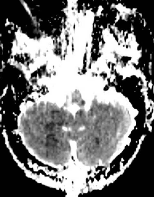
This is a case of multiple embolic infarcts following a diagnostic four vessel angiogram for a left internal cerebral artery aneurysm following clipping in a 72-year-old female. The first image is a FLAIR sequence demonstrating several foci of hyperintense signal involving the deep and subcortical white matter as well as the cortical grey matter of the left frontal lobe. The second and third images are the corresponding DWI and ADC maps demonstrating several foci of restricted diffusion at the corresponding sites of FLAIR hyperintensity. The next image demonstrates several foci of FLAIR hyperintense signal involving the cerebellar hemispheres, predominantly on the right, and the right cerebellar tonsil. The last two images are the corresponding DWI and ADC maps eliciting several foci of diffusion restriction on the right. Thromboembolic events are a principle cause of ischemic stroke. Emboli may arrise from arterial stenoses or occlusions, right to left shunts in the setting of deep venous thrombosis, or cardiac sources. Cardiac sources account for approximately 15 to 20% of ischemic strokes.
Related videos to the case