- 2
- ,
- 2
- 4
- 6
To Quiz Yourself: Select OFF by clicking the button to hide the diagnosis & additional resources under the case.
Quick Browser: Select ON by clicking the button to hide the additional resources for faster case review.
CASE NUMBER
124


Diagnosis
Developmental Venous Anomaly
Note
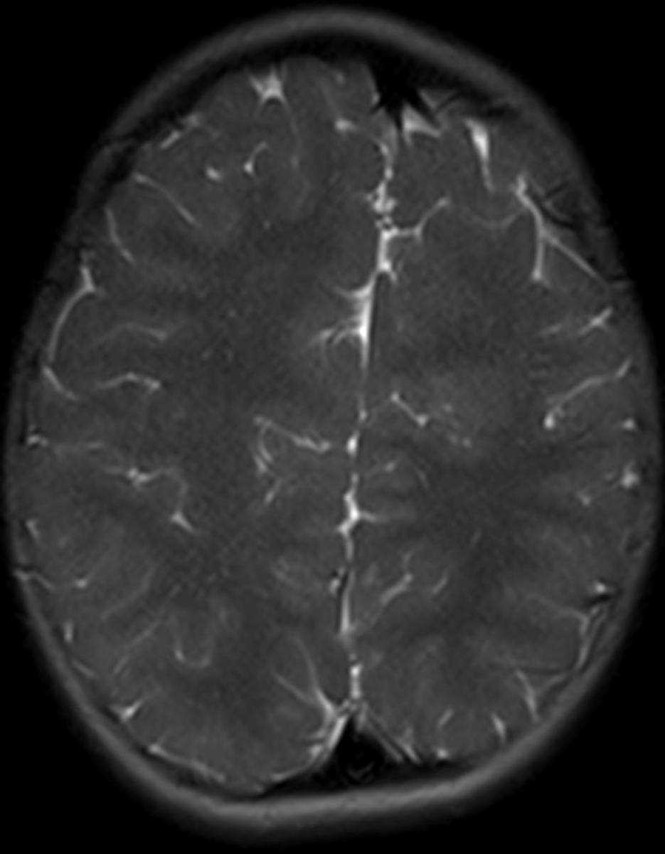
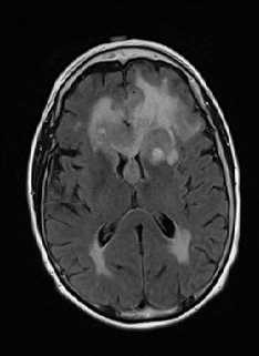
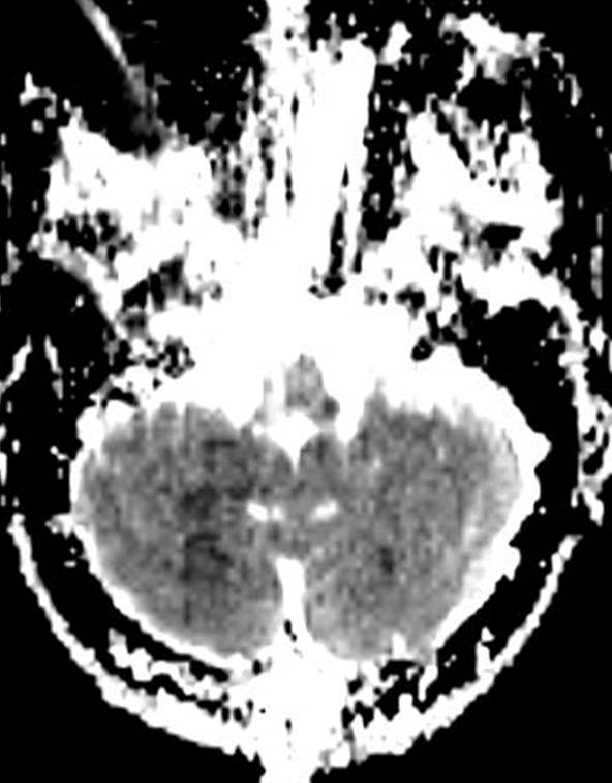
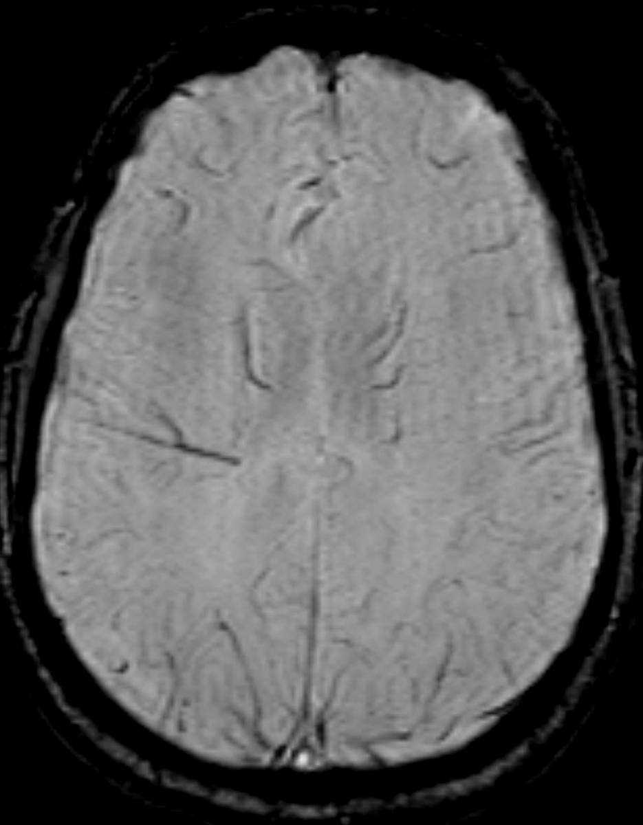
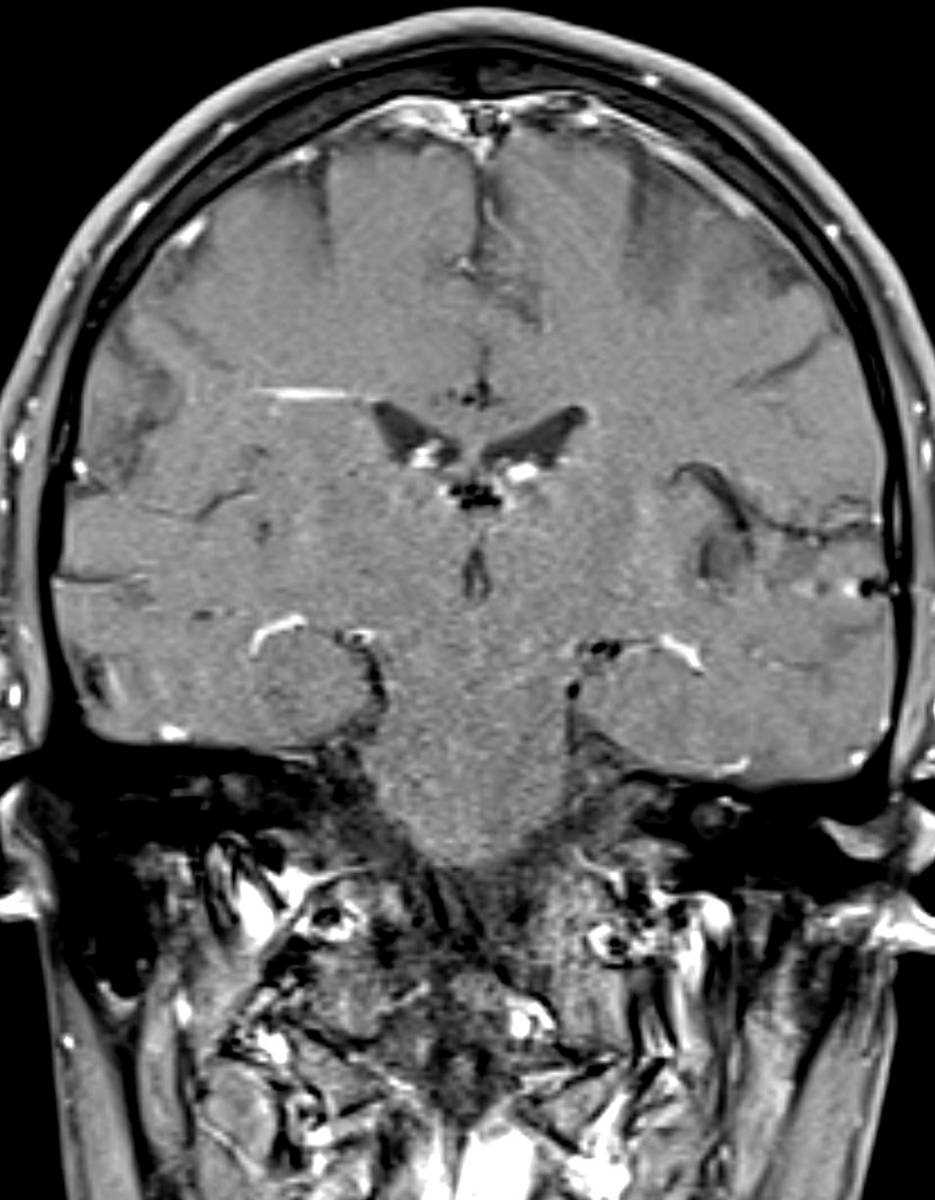
This is a case of an incidentally detected developmental venous anomaly, or DVA, on a follow-up brain MR for an arachnoid cyst in a 38-year-old female. The first two images demonstrate the linear hypointense lesion coursing through the deep and periventricular white matter of the right frontoparietal region on the susceptibility weighted sequences. The third image, which is a T2-weighted sequence, demonstrates a corresponding linear T2 hyperintense signal at the DVA. The fourth and fifth images demonstrate the DVA on post contrast T1-weighted sequences with linear enhancement and extension towards the subependymal veins on the right. Two middy dilated subependymal veins are demonstrated on the right on the final postcontrast image. A DVA results from a network of dilated medullary veins radially converging on a large vein that drains into deep or superficial veins. A DVA is surrounded by normal brain parenchyma. DVAs are thought to arrise from prenatal occlusion of a vein draining into the deep or superficial venous system. This results in a large collateral vein forming which drains through the white matter. Because the DVA represents a compensatory venous drainage pathway for normal brain, sacrifice of this pathway can result in venous infarction.
Related videos to the case

THIS IS CASE
124
OF
373