- 2
- ,
- 2
- 4
- 6
To Quiz Yourself: Select OFF by clicking the button to hide the diagnosis & additional resources under the case.
Quick Browser: Select ON by clicking the button to hide the additional resources for faster case review.
CASE NUMBER
122


Diagnosis
Primary CNS Lymphoma
Note
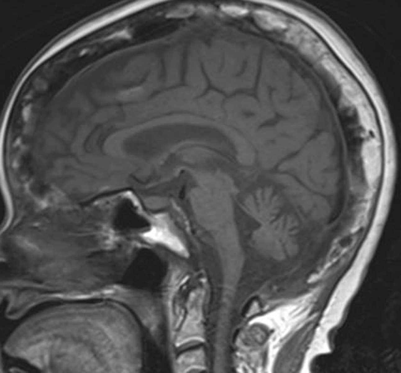
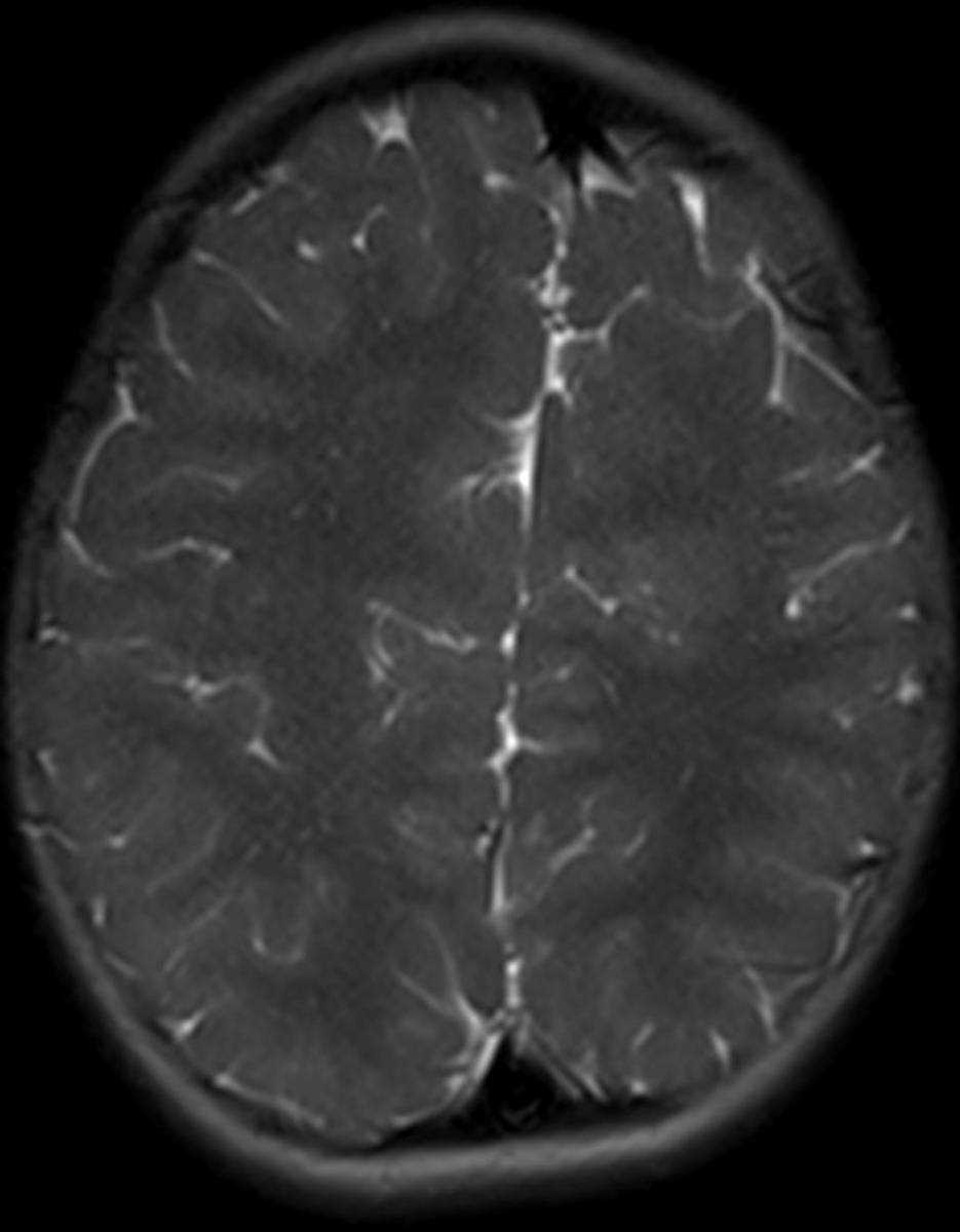
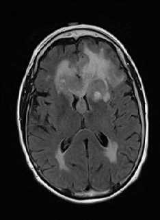
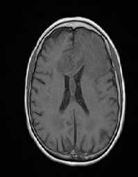
This is a case of primary CNS lymphoma in a 54-year-old female presenting with new onset seizures. The first image demonstrates a confluent FLAIR hyperintense lesion predominantly centered within the deep gray matter of the left basal ganglia and extending along the periventricular white matter surfaces. The second image, which is a non-contrast T1-weighted image and third through fourth images, which are post contrast T1-weighted sequences, demonstrate marked peripheral enhancement of the centrally necrotic lesion centered in the left periventricular white matter. In addition, there is involvement of and extension along the corpus callosum. The fifth and sixth images demonstrate diffusion restriction with corresponding hyperintensity and hypointensity on the DWI and ADC maps, respectively. CNS lymphoma is often associated with an immunodeficiency state, such as AIDS, organ transplantation, or collagen vascular diseases where immunomodulators are commonly used in therapy. Depending on the patients immune status and underlying chronic medical disorders, the appearance is variable with multiple necrotic masses, ventriculitis, CSF infiltration or a single necrotic mass a possibility.
Related videos to the case


THIS IS CASE
122
OF
373