- 2
- ,
- 3
- 2
- 7
To Quiz Yourself: Select OFF by clicking the button to hide the diagnosis & additional resources under the case.
Quick Browser: Select ON by clicking the button to hide the additional resources for faster case review.
CASE NUMBER
314


Diagnosis
Metastatic Pancreatic Adenocarcinoma
Note
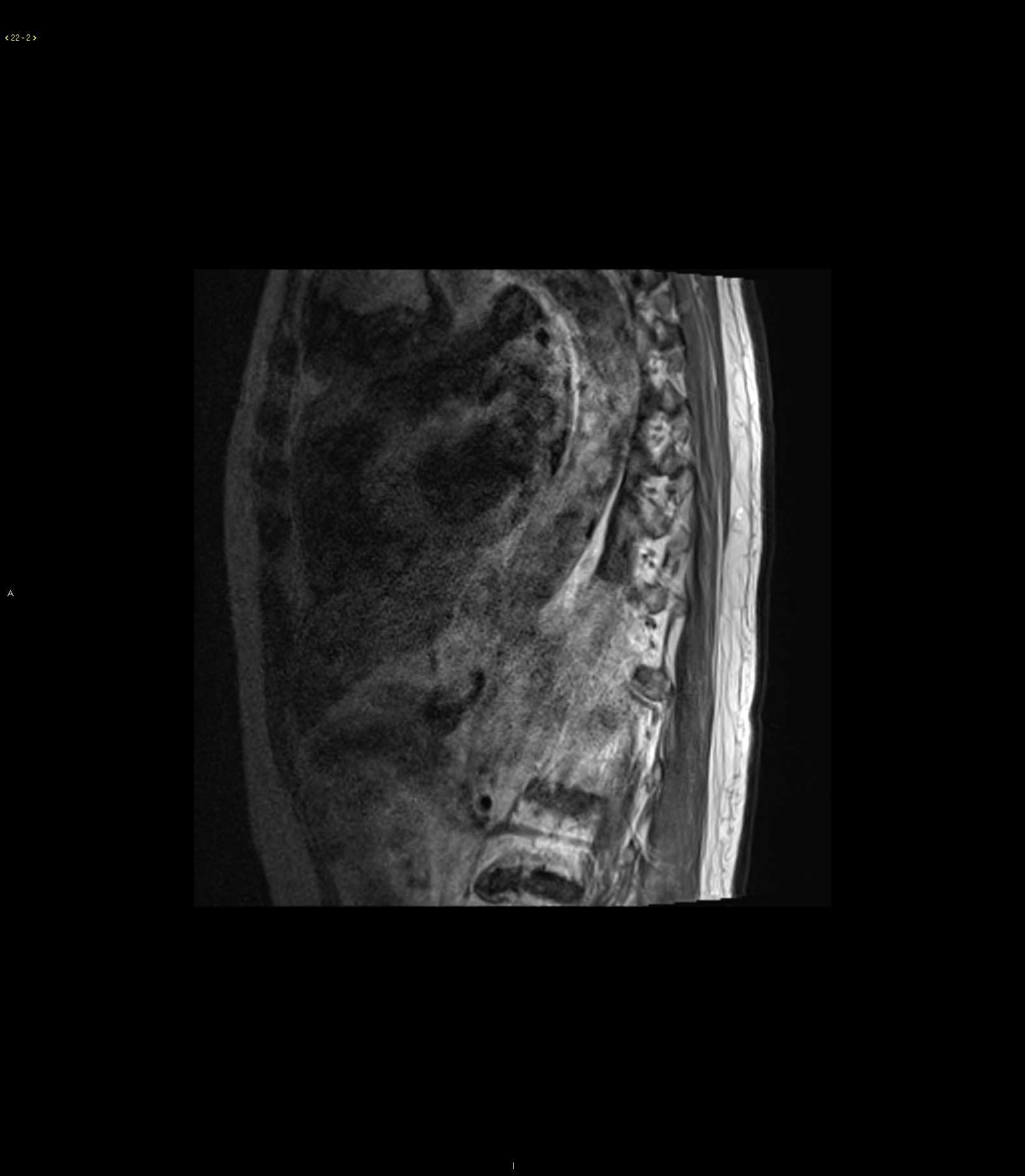
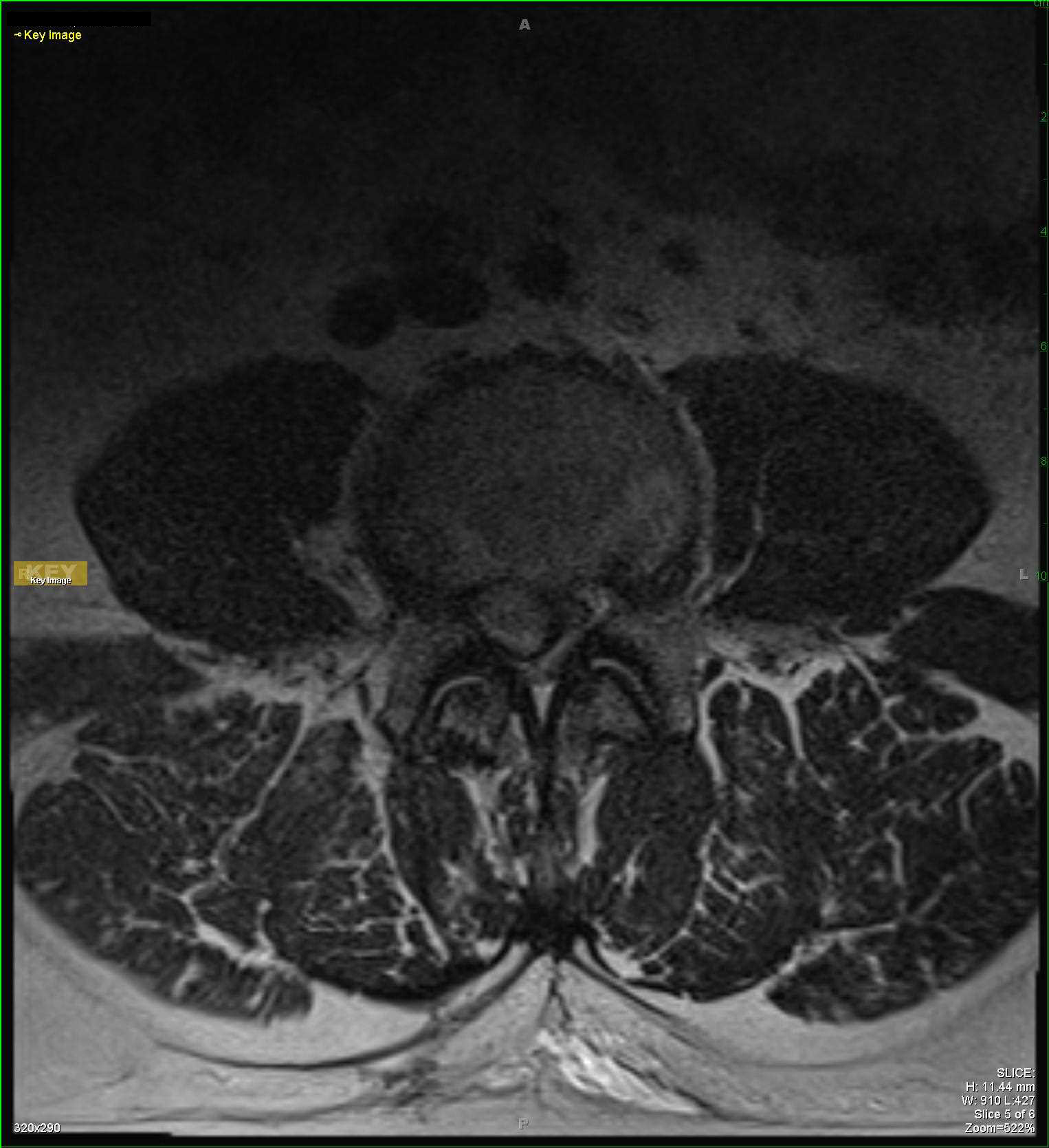
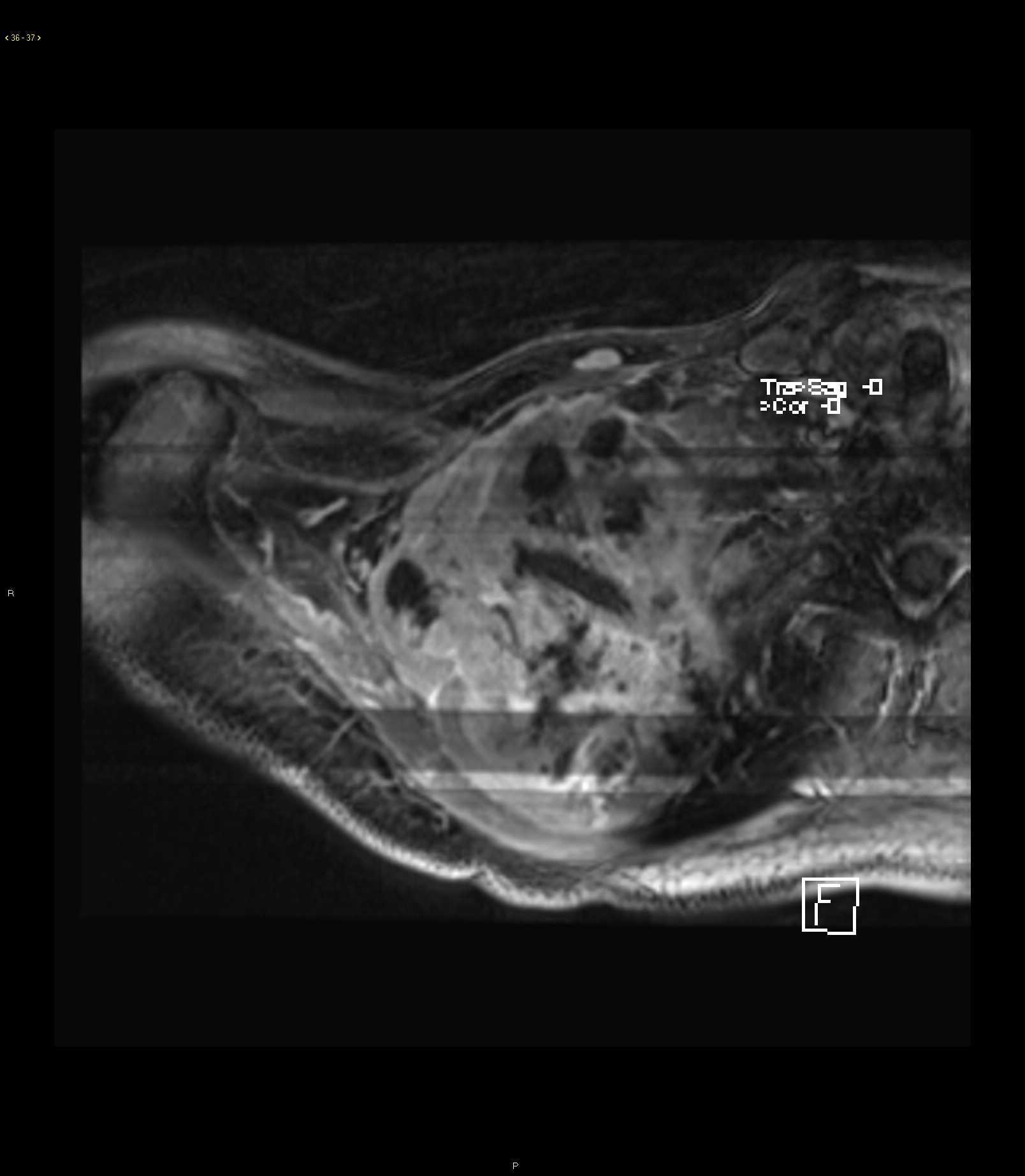
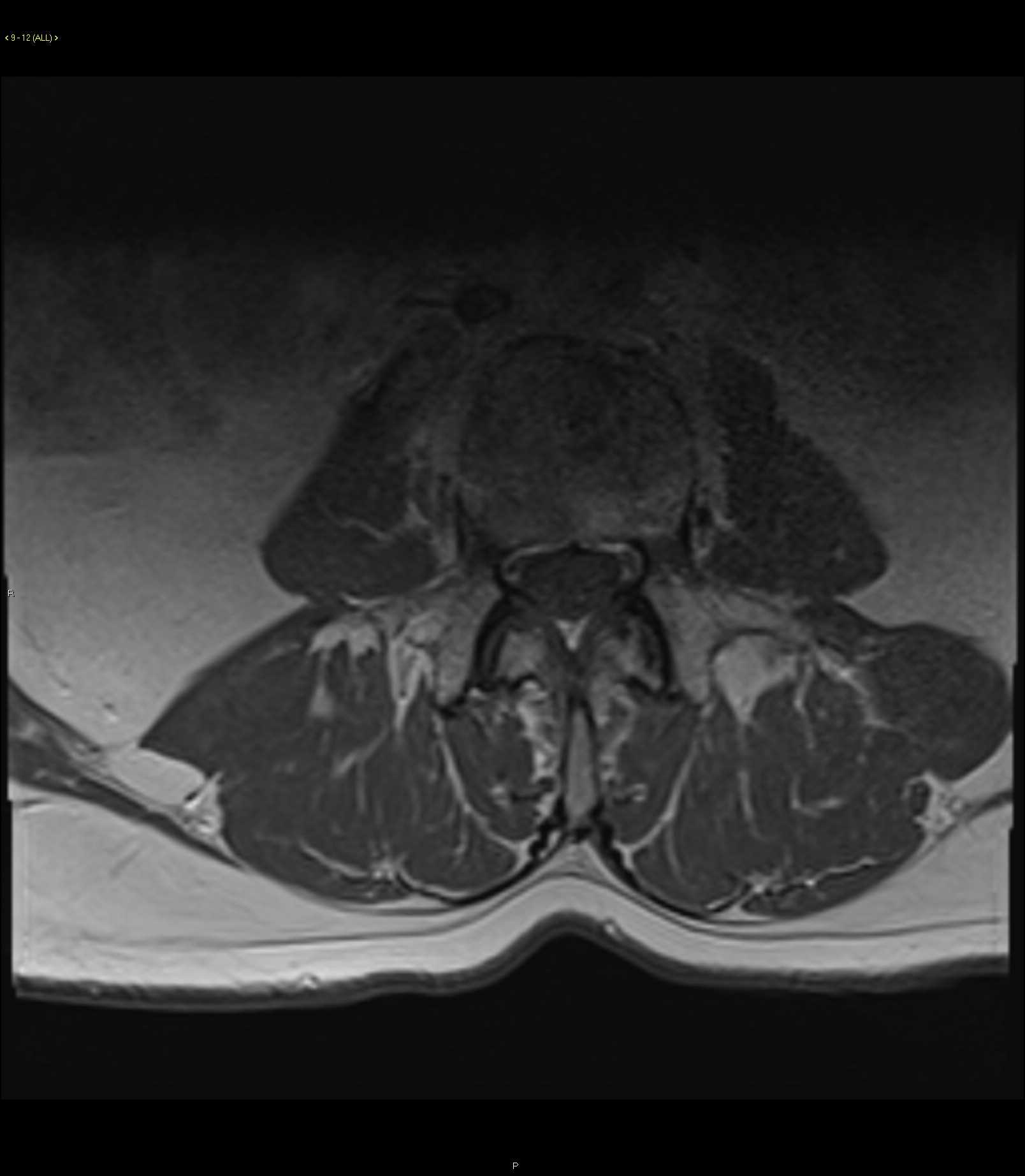
49-year-old male with history of pancreatic adenocarcinoma status post Whipple followed by radiation therapy. The images demonstrate an infiltrative T1 hypointense, T2 heterogeneously hypointense, STIR heterogeneously hyperintense mass involving the L3 vertebral body. The axial images demonstrate an infiltrative marrow lesion extending into the pedicle posteriorly on the right. The lesion demonstrates heterogeneous enhancement with an associated soft tissue component extending along the anterior lateral margins of the vertebral body. Imaging findings are most compatible with osseous metastatic disease with secondary extension into the adjacent soft tissues at the L3 vertebral level. Osseous metastatic disease accounts for over one half of all malignant bone tumors. Lung cancer, breast cancer, renal cell carcinoma and prostate cancer account for greater than three quarters of all osseous metastatic disease. The majority of osseous metastatic lesions developed from hematogenous spread. Distribution of skeletal metastatic disease mimics the distribution of red marrow.
Related videos to the case