- 2
- ,
- 3
- 2
- 7
To Quiz Yourself: Select OFF by clicking the button to hide the diagnosis & additional resources under the case.
Quick Browser: Select ON by clicking the button to hide the additional resources for faster case review.
CASE NUMBER
313


Diagnosis
Malignant Mediastinal Teratoma
Note
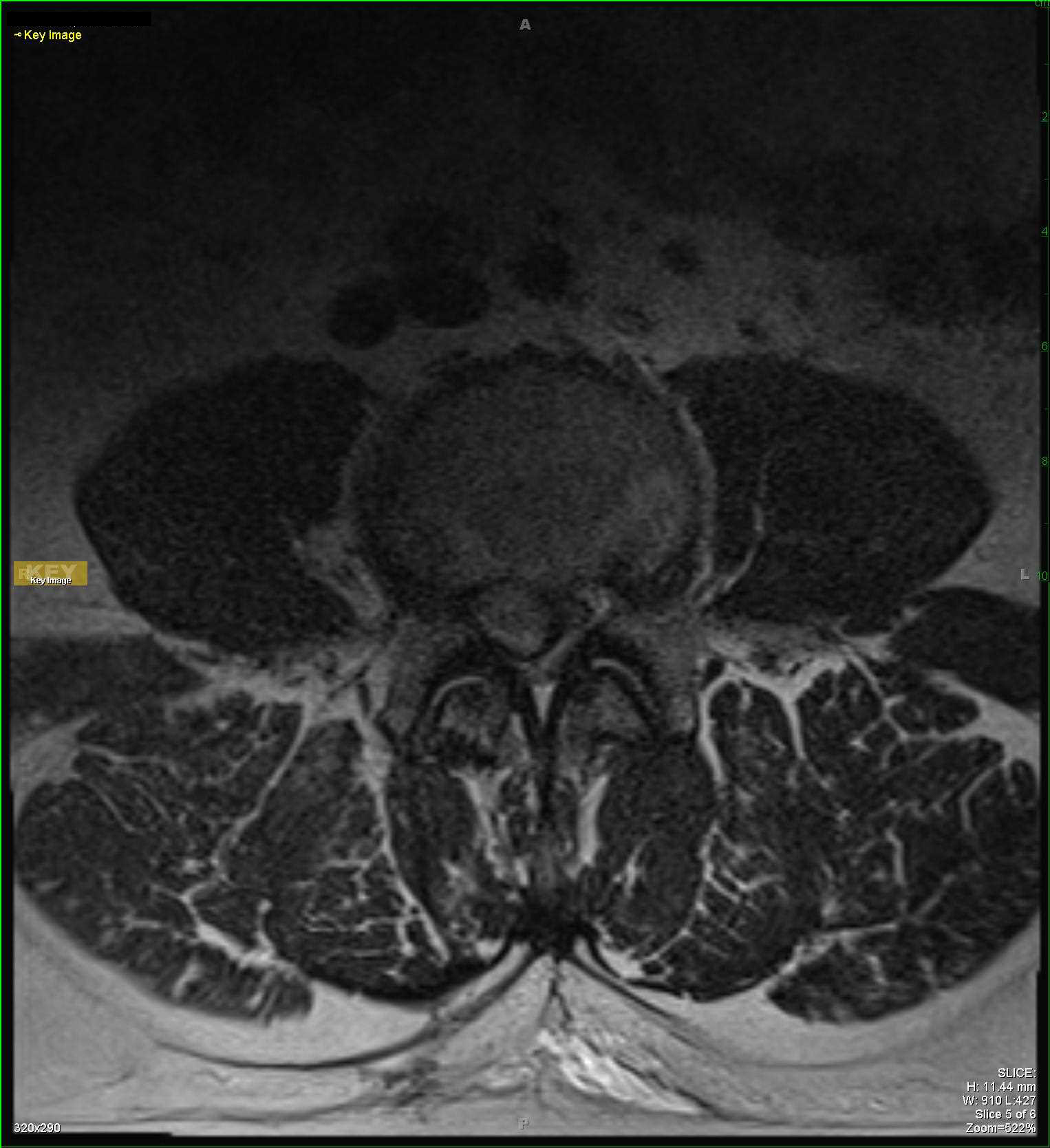
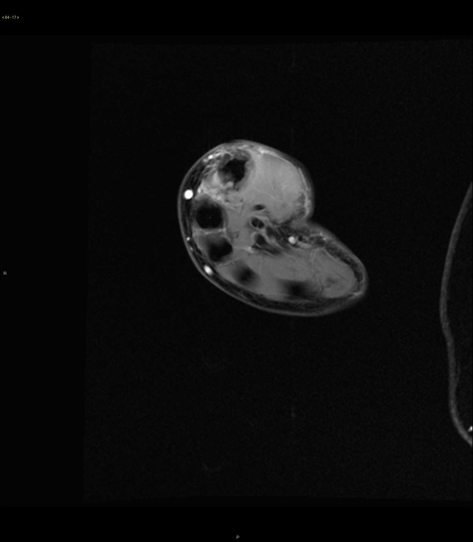
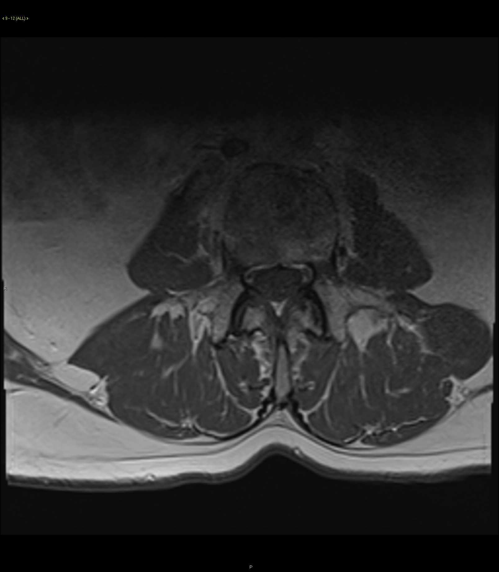
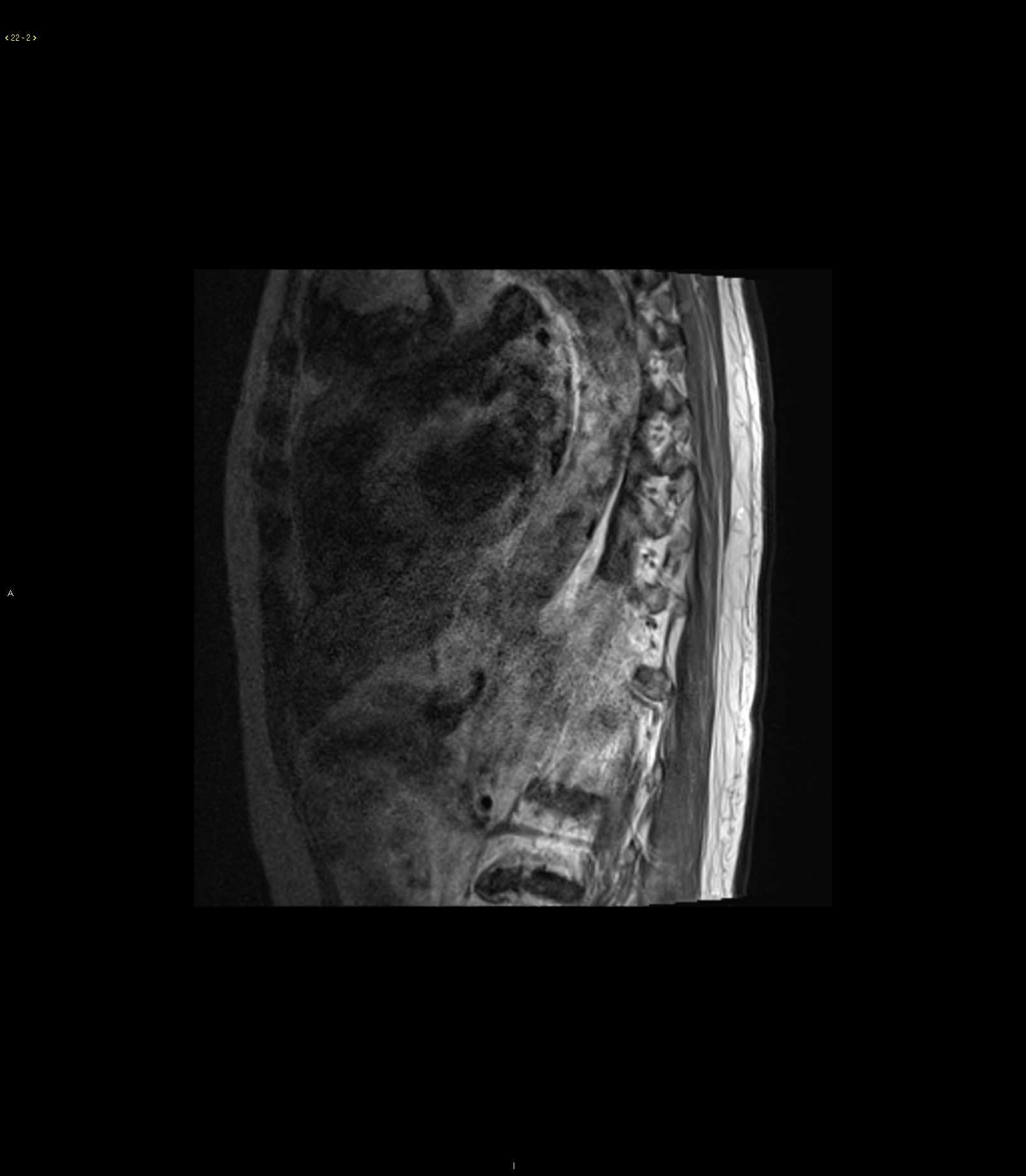
63-year-old female with history of low back pain. There is a solid and cystic appearing partially circumscribed anterior mediastinal mass with components that are both T2 hyper- and hypointense as well as T1 hyper- and hypointense. On the fat saturated postcontrast images, there is a rind of enhancing tissue with a more solid enhancing mural component at its inferior most aspect. There is also a single lower thoracic vertebral body which demonstrates a T1 hypointense, T2/STIR hyperintense lesion arising from its inferior endplate with extension towards the pedicle. Imaging findings are most compatible with malignant degeneration of anterior mediastinal mature teratoma. Mediastinal teratomas account for less than one quarter of all anterior mediastinal lesions in adults. They are the most common extragonadal germ cell tumor, accounting for over one half of all mediastinal germ cell tumors. Typical age at presentation in adults is in the third to fourth decade of life. Clinical presentation is usually due to either mass effect, endocrine function, or rupture. Solid lesions tend to be malignant while cystic lesions tend to be more benign.
Related videos to the case


THIS IS CASE
313
OF
370