- 2
- ,
- 3
- 8
- 1
To Quiz Yourself: Select OFF by clicking the button to hide the diagnosis & additional resources under the case.
Quick Browser: Select ON by clicking the button to hide the additional resources for faster case review.
CASE NUMBER
381


Diagnosis
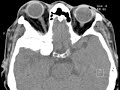
Sphenoid Wing Meningioma
Note
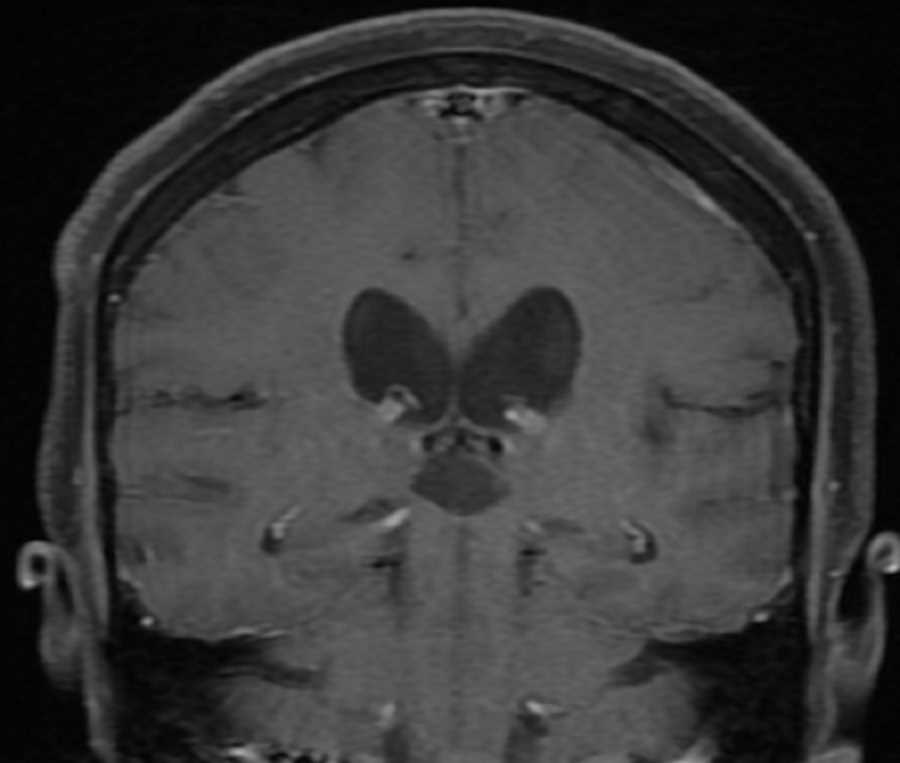
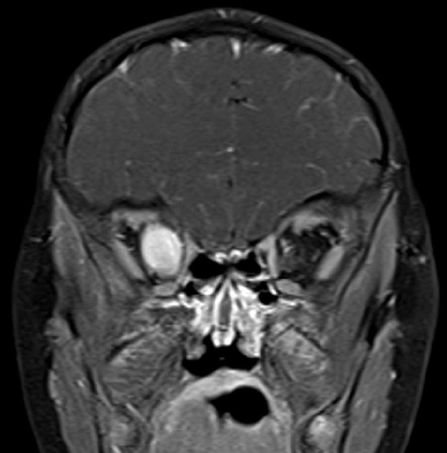
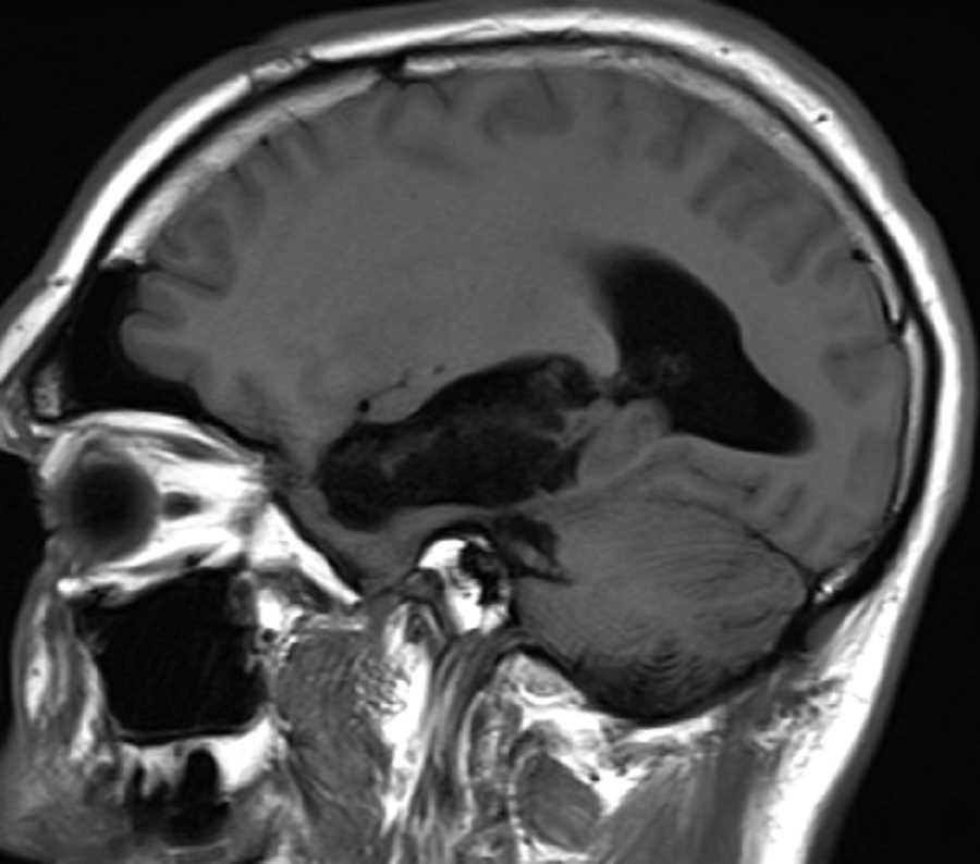
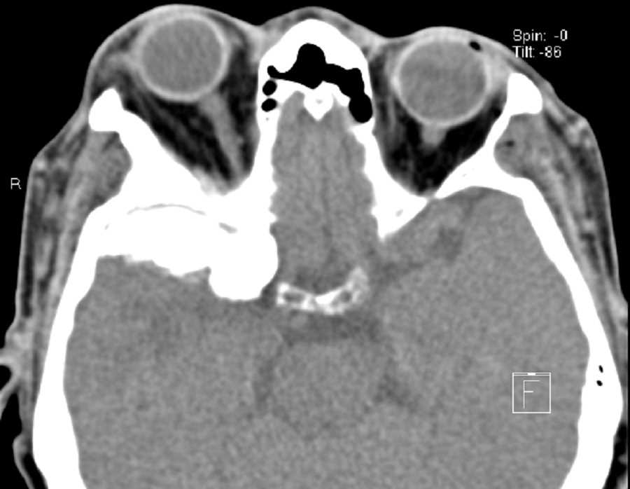
These images demonstrate a hyperdense infiltrative mass located in the anterior right middle cranial fossa with impressive reactive hyperostosis involving the greater sphenoid wing with extension into the right orbital apex, skull base foramina, and right sphenoid sinus. There is hypoattenuation in the anterior right temporal lobe likely reflecting gliosis. This mass was favored to represent a meningioma, although metastasis and primary bone tumors were also considered in the differential. Meningiomas are the most common primary intracranial tumor, are more common in females, usually appear hyperdense on CT, and may calcify. They usually avidly enhance and can have reactive hyperostosis of the adjacent calvarium as seen here.
Related videos to the case

THIS IS CASE
381
OF
396