- 2
- ,
- 2
- 4
- 6
To Quiz Yourself: Select OFF by clicking the button to hide the diagnosis & additional resources under the case.
Quick Browser: Select ON by clicking the button to hide the additional resources for faster case review.
CASE NUMBER
357


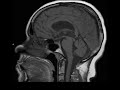
Diagnosis
Pineal Gland Cyst
Note
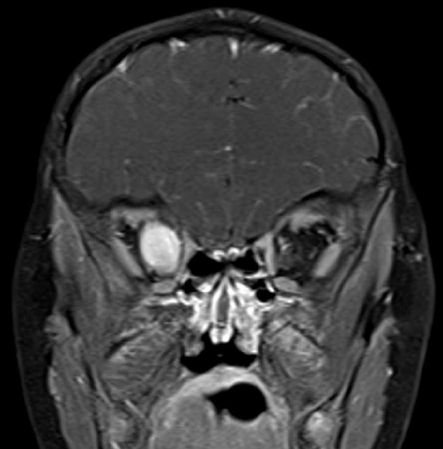
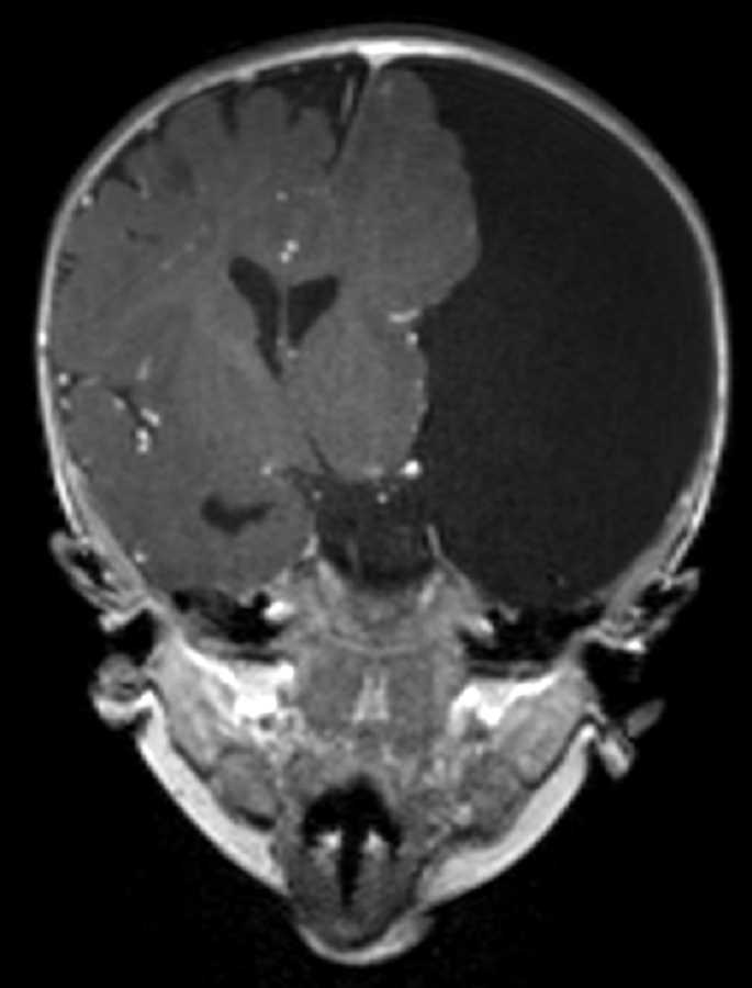
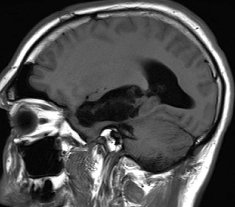
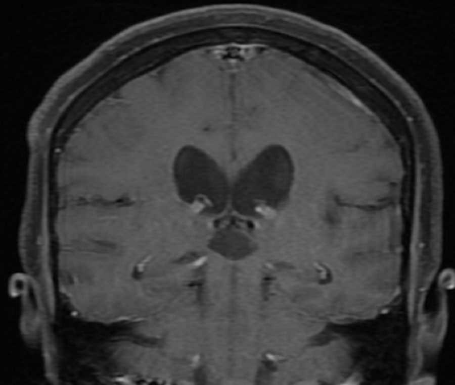
These images show a T1 hypointense, T2 hyperintense lesion in the posterior aspect of the third ventricle which appears to arise from the anterior aspect of the pineal gland. This lesion does not restrict diffusion or enhance and incomplete suppression on T2 FLAIR. The mass results in mild mass effect on the anterior aspect of the tectal plate and subsequently the cerebral aqueduct. There is mild supratentorial ventriculomegaly, but no evidence of transependymal flow as would be seen in acute obstructive hydrocephalus. Symptoms of a lesion in this location include Parinaud syndrome (vertical gaze palsy). Findings are most consistent with a pineal gland cyst. Differential considerations include pineocytoma (usually has solid enhancing components, but imaging features may overlap), arachnoid cyst (follows CSF on sequences), and epidermoid (should restrict diffusion).
Related videos to the case

THIS IS CASE
357
OF
373