- 2
- ,
- 2
- 4
- 6
To Quiz Yourself: Select OFF by clicking the button to hide the diagnosis & additional resources under the case.
Quick Browser: Select ON by clicking the button to hide the additional resources for faster case review.
CASE NUMBER
352


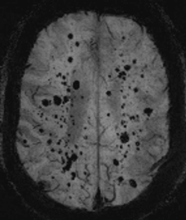
Diagnosis
Multiple Cavernoma Syndrome
Note
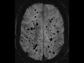
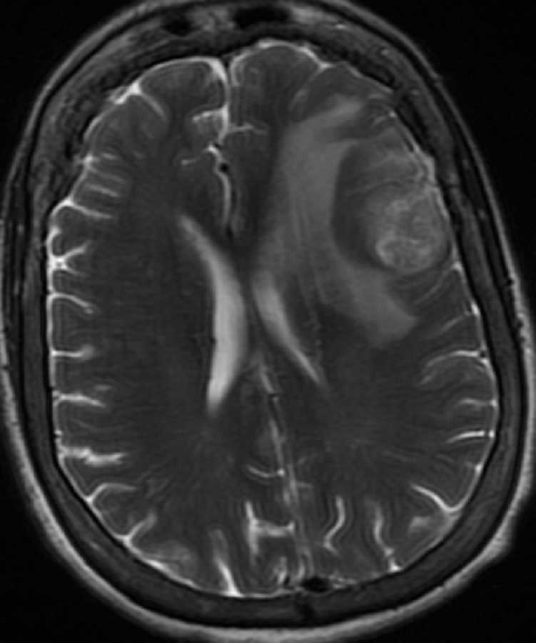
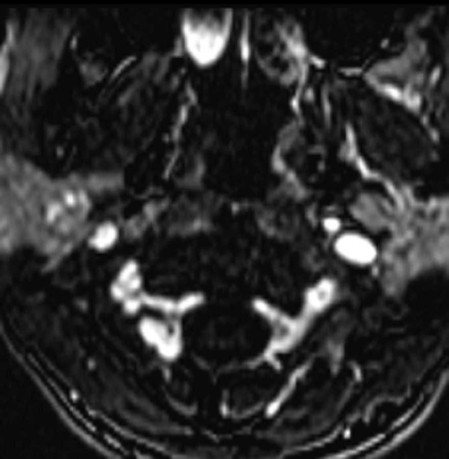
There are innumerable foci of increased susceptibility through both cerebral hemispheres, the cerebellum, and the brainstem. The largest lesion located in the periventricular white matter of the left frontal lobe has a popcorn appearance with peripheral areas of intrinsic T1 hyperintensity compatible with intralesional hemorrhage. The differential diagnosis includes multiple cavernoma syndrome, amyloid angiopathy, and microhemorrhages related to diffuse axonal injury. Given the brainstem involvement and the classic appearance of the lesion in the left frontal lobe for cavernoma, the best diagnosis in the case is multiple cavernoma syndrome. This is an autosomal dominant syndrome with variable penetrance. The mean bleeding rate is close to 1% per lesion per year. Cavernoma formation is also associated with brain radiation usually received in childhood with an average latency period of approximately 20 years.
Related videos to the case