- 2
- ,
- 2
- 5
- 2
To Quiz Yourself: Select OFF by clicking the button to hide the diagnosis & additional resources under the case.
Quick Browser: Select ON by clicking the button to hide the additional resources for faster case review.
CASE NUMBER
308


Diagnosis
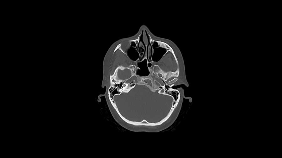
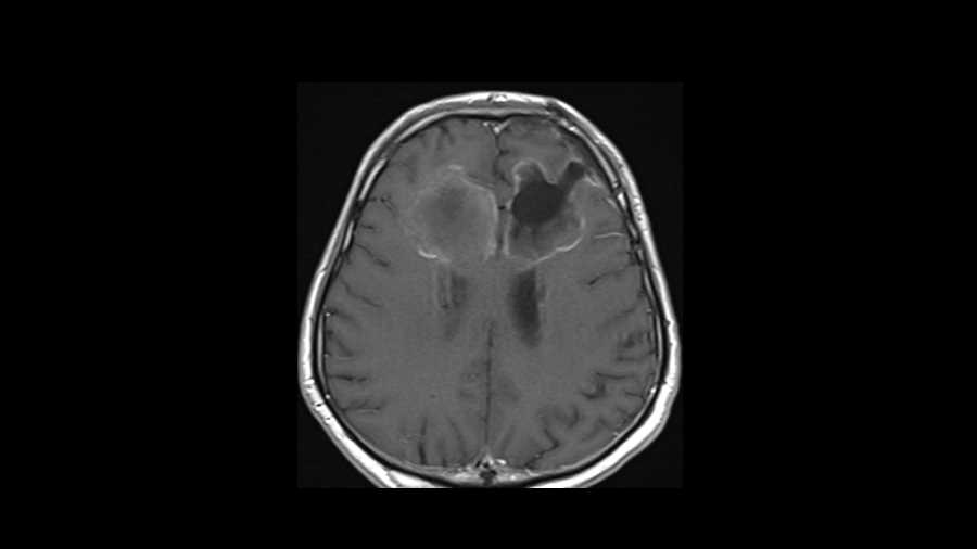
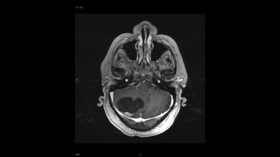
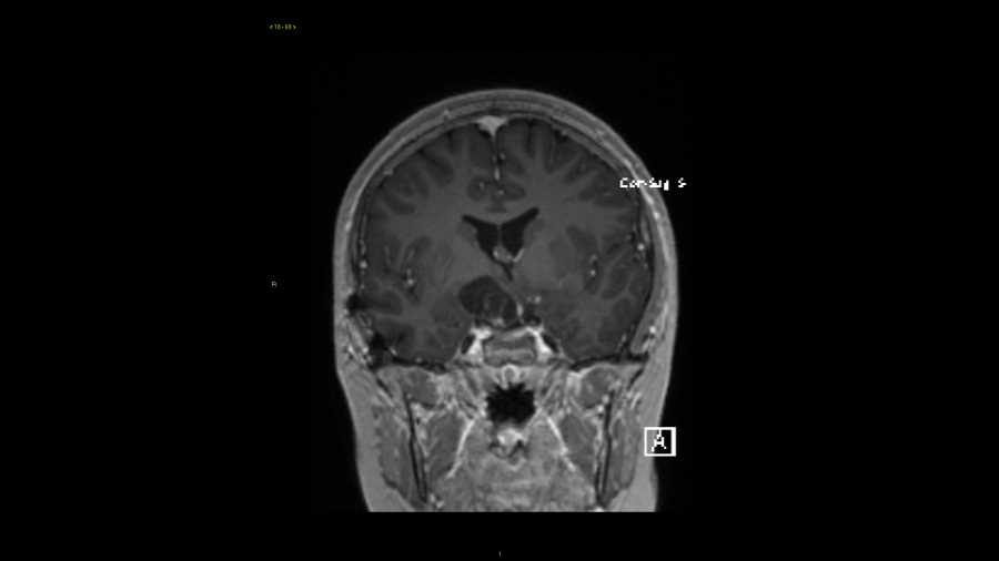
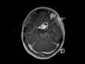
Optic nerve glioma
Note
These images show a 2 cm lobulated T1 hypointense, T2 FLAIR hyperintense mass in the suprasellar cistern with cystic and enhancing solid components. There is mild mass effect on the superior aspect of the pituitary and the infundibulum is displaced to the left. The optic chiasm is obliterated by the mass and there is enlargement of the prechiasmatic left optic nerve. This turned out to be optic nerve glioma in a patient with a history of NF-1. Without the optic nerve involvement in a young child, the differential considerations include craniopharyngioma or germ cell tumor.
Related videos to the case

THIS IS CASE
308
OF
374