- 2
- ,
- 2
- 4
- 6
To Quiz Yourself: Select OFF by clicking the button to hide the diagnosis & additional resources under the case.
Quick Browser: Select ON by clicking the button to hide the additional resources for faster case review.
CASE NUMBER
273


Diagnosis
Solitary Fibrous Tumor of the Right Orbit
Note
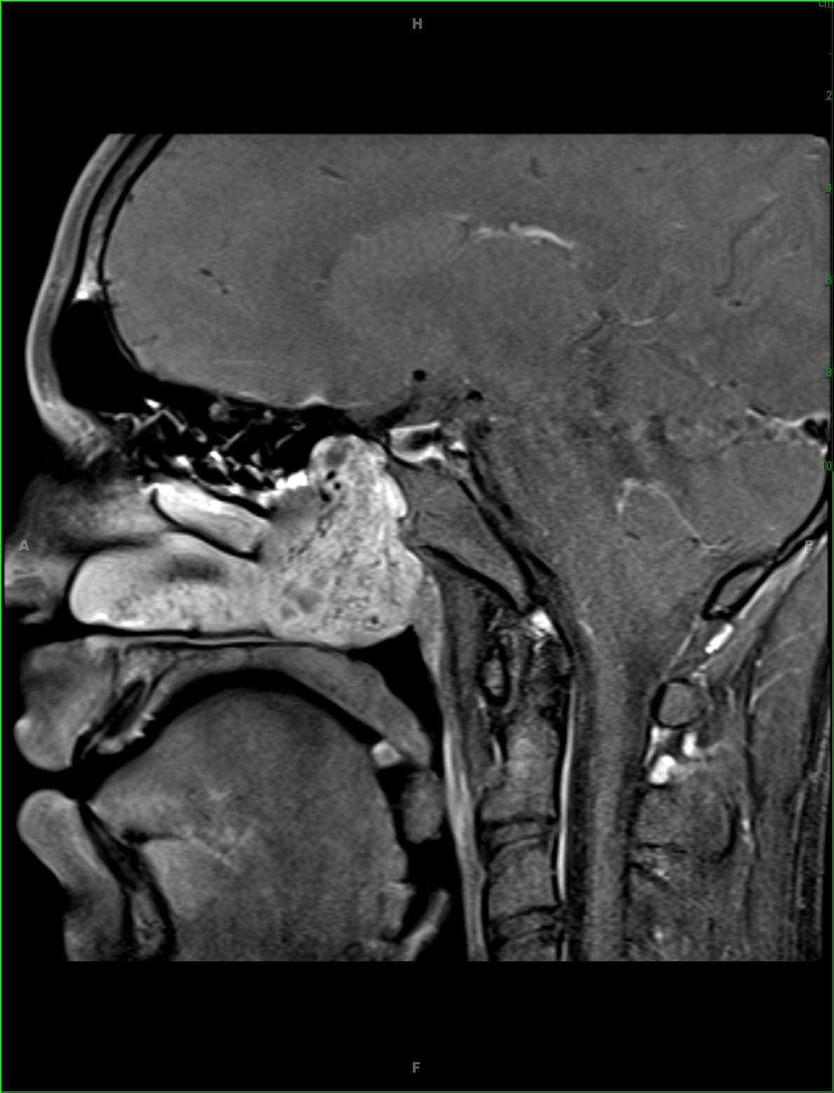
52-year-old female with history of slow onset painful proptosis on the right. There is an infiltrative poorly circumscribed T1 hypointense T2 hyperintense, heterogeneously enhancing mass centered at the posterior aspect of the right orbit. There is evidence of chronic osseous remodeling along the roof of the orbital the right with infiltrative enhancing tissue and dural thickening extending posteriorly through the region of the orbital apex to the optic canal and superior oblique fissure on the right. On biopsy, this lesion was found to be a solitary fibrous tumor of the orbit. Solitary fibrous tumor is an uncommon spindle-shaped cell tumor of mesenchymal origin. Lesions typically arise in the pleura, but head and neck lesions may be seen in the sinonasal cavity, nasopharynx, larynx, parapharyngeal space, floor of the mouth, thyroid and orbits. MRI is the best technique for mapping the extent of the lesion and relationship to surrounding structures. The differential for such a lesion may include orbital schwannoma, hemangiopericytoma, pseudotumor or lacrimal gland tumor. Treatment is with complete surgical excision.
Related videos to the case