- 2
- ,
- 3
- 8
- 1
To Quiz Yourself: Select OFF by clicking the button to hide the diagnosis & additional resources under the case.
Quick Browser: Select ON by clicking the button to hide the additional resources for faster case review.
CASE NUMBER
274
Diagnosis
Grade 3 Anaplastic Astrocytoma
Note
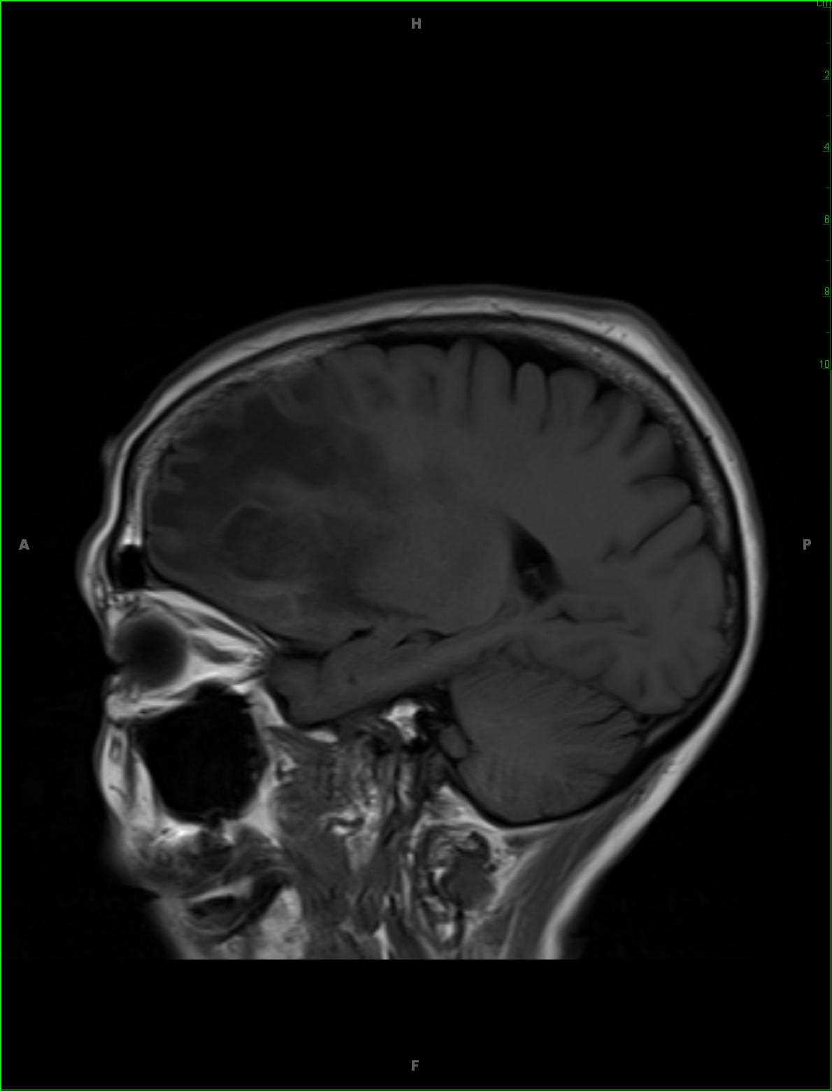
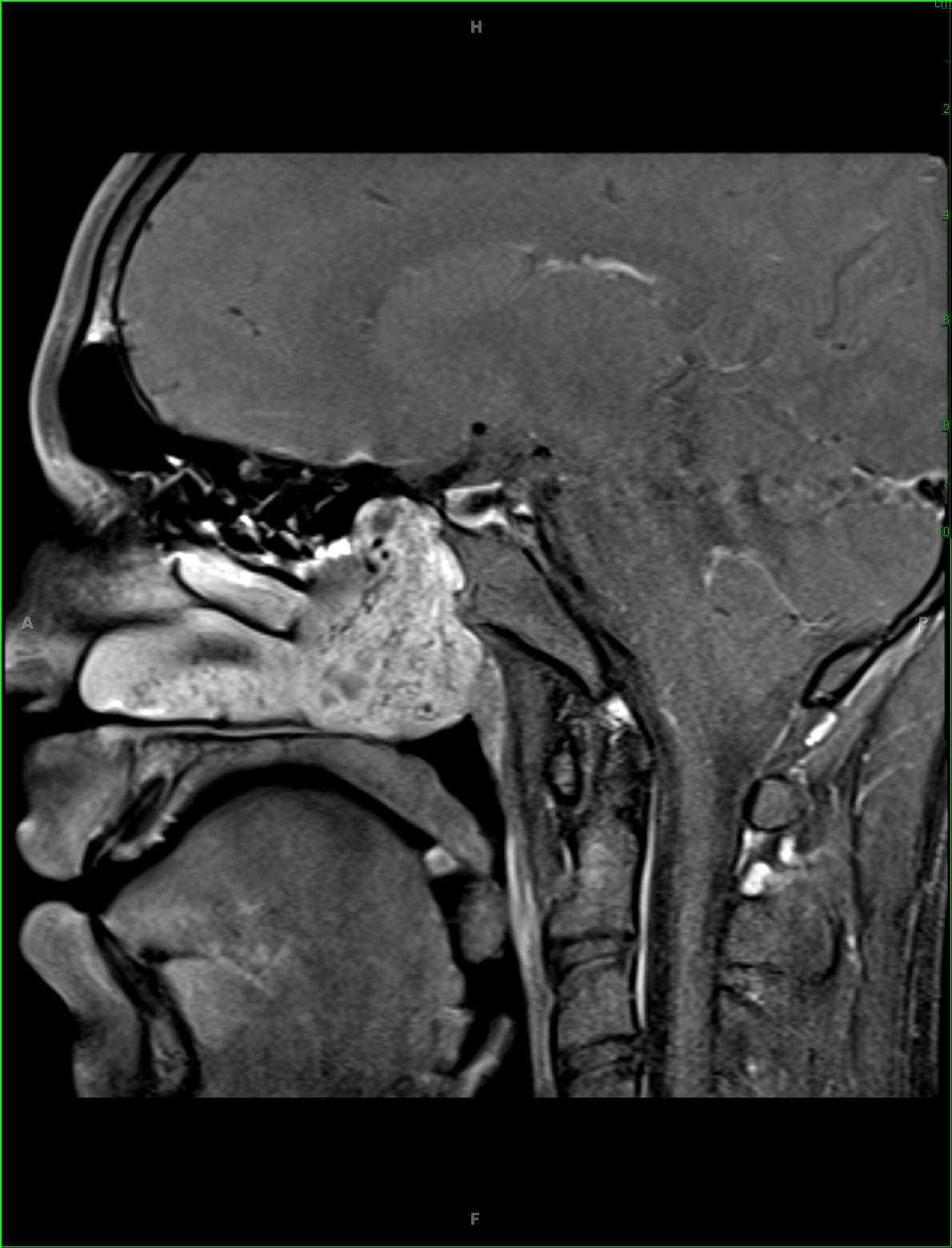
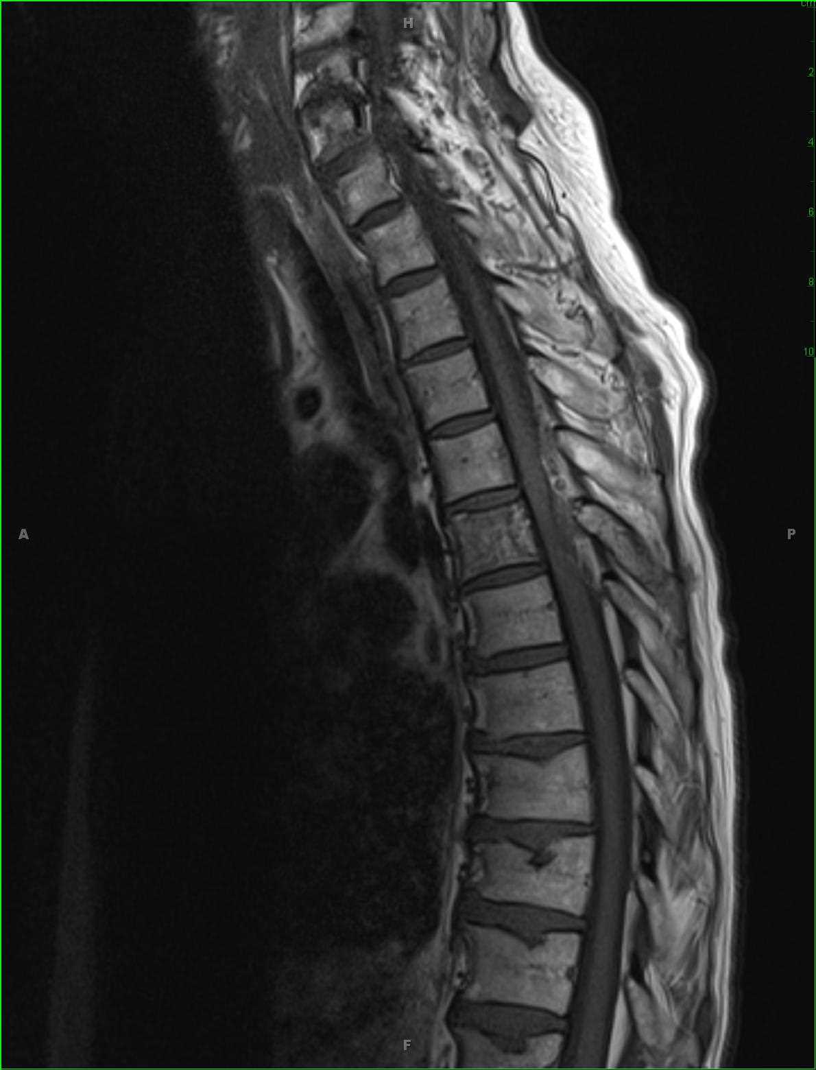
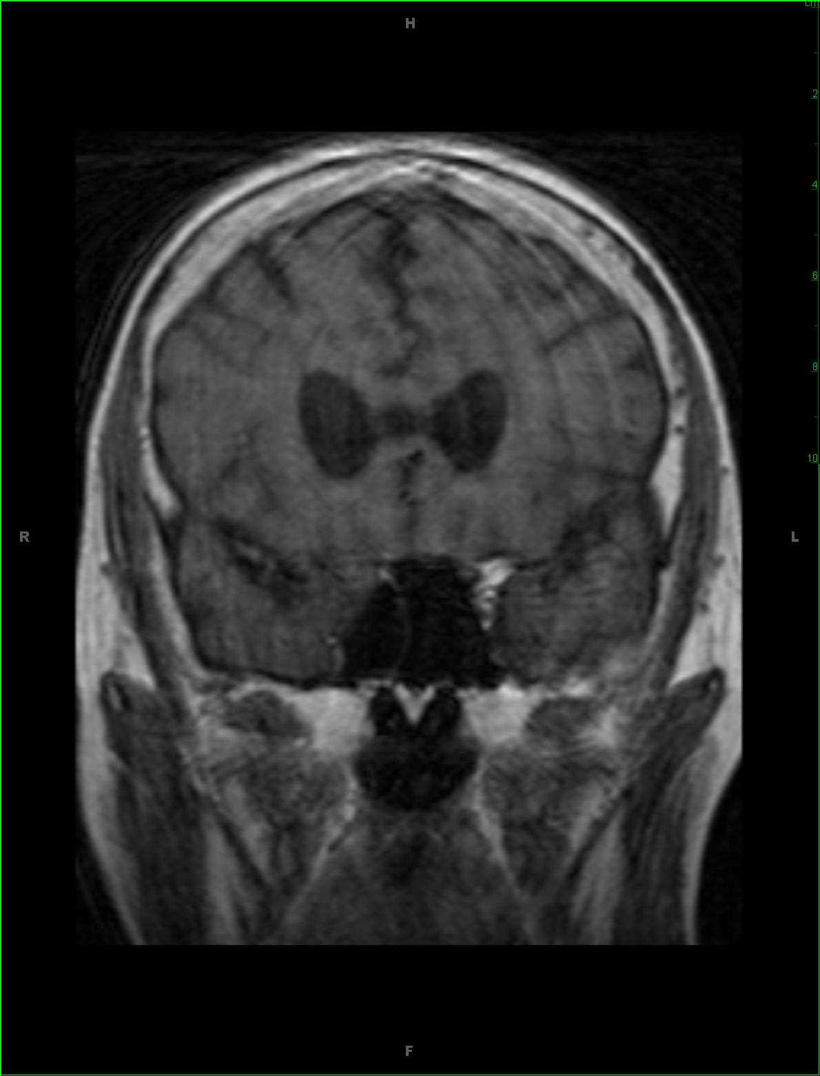
58-year-old male presenting for chronic headaches. There is an infiltrative T1 hypointense, T2/FLAIR hyperintense mass centered within the periventricular deep white matter of the left frontal region with extension to the insula and corpus striatum on the left. There is T2 hyperintense signal which extends across the genu and body of the corpus callosum. The lesion results in partial effacement of the frontal horn and body of the left lateral ventricle with slight rightward deviation of the septum pellucidum. There are irregular regions of low ADC values within the central periventricular component without evidence of increased blood volume on the perfusion weighted images. The lesion is demonstrates nodular enhancing components centrally. The differential includes high-grade glioma, lymphoma, and metastatic disease. On biopsy, the lesion was deemed to be a grade 3 anaplastic astrocytoma. Anaplastic astrocytoma is considered a WHO Grade III tumor. These lesions usually occur in adulthood, with peak incidence between 40 and 50 years. Whereas diffuse low grade astrocytoma will not demonstrate enhancement, anaplastic astrocytoma will lack regions of frank necrosis, a finding seen in glioblastoma, WHO Grade IV tumors.
THIS IS CASE
274
OF
396


