- 2
- ,
- 2
- 4
- 6
To Quiz Yourself: Select OFF by clicking the button to hide the diagnosis & additional resources under the case.
Quick Browser: Select ON by clicking the button to hide the additional resources for faster case review.
CASE NUMBER
255


Diagnosis
Tuberous Sclerosis
Note
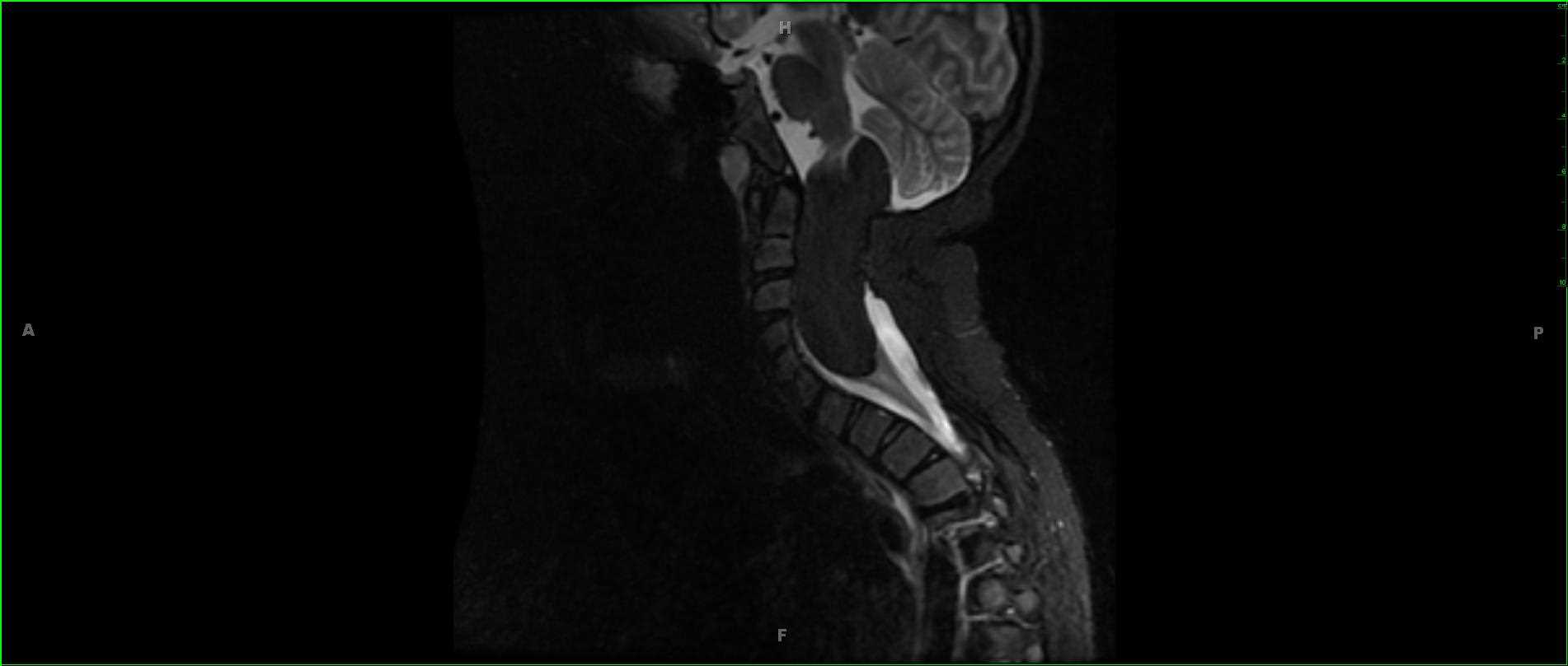
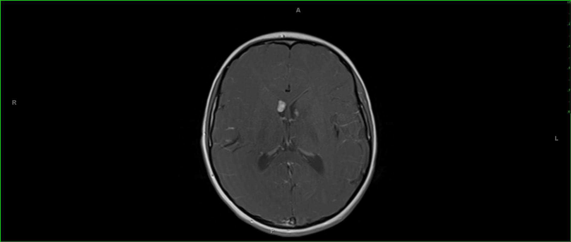
11-year-old male with chronic seizures. The axial T1-weighted images demonstrate nodular regions of isointense signal to deep white matter along the lateral ventricular margins at the level of the frontal horns. A sagittal off midline T1-weighted image redemonstrates the nodular regions of T1 isointense signal along the ventricular margins as well as a small focal region of encephalomalacia in the body of the corpus callosum. There are linear, bandlike, T2/FLAIR hyperintense regions in the subcortical and deep white matter of the cerebral hemispheres, right greater than left. Scattered regions of susceptibility are identified in the sub- and juxtacortical white matter of both the right and left cerebral hemispheres. Postcontrast images demonstrate nodular regions of enhancement in the subcortical white matter as well as enhancing nodules along the ependymal margins of the frontal horns of the lateral ventricles. The spectrum of findings are classic for tuberous sclerosis. Tuberous sclerosis falls in the category of phakomatosis or neuro-oculocutaneous syndromes which include neurofibromatosis, ataxia telengectasia, Sturge-Weber syndrome, von Hippel-Lindau disease and Nevoid basal cell carcinoma syndrome, among others. Clinical presentation typically includes seizures, mental retardation, and adenoma sebaceum. Tuberous sclerosis results from mutations in tumor suppressor genes, TSC1 which is on chromosome 9 and TSC2, chromosome 16.
Related videos to the case

THIS IS CASE
255
OF
373