- 2
- ,
- 2
- 4
- 6
To Quiz Yourself: Select OFF by clicking the button to hide the diagnosis & additional resources under the case.
Quick Browser: Select ON by clicking the button to hide the additional resources for faster case review.
CASE NUMBER
254


Diagnosis
Synovial Sarcoma, Foot
Note
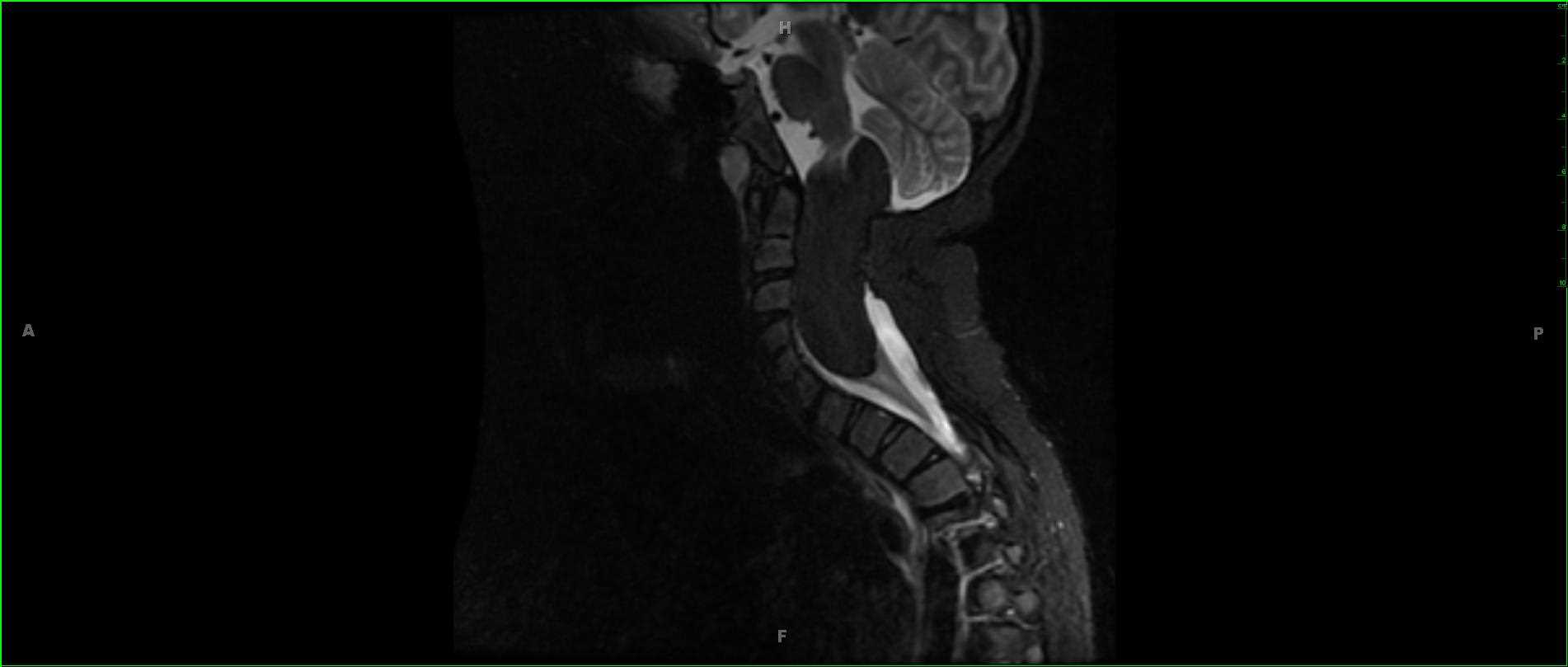
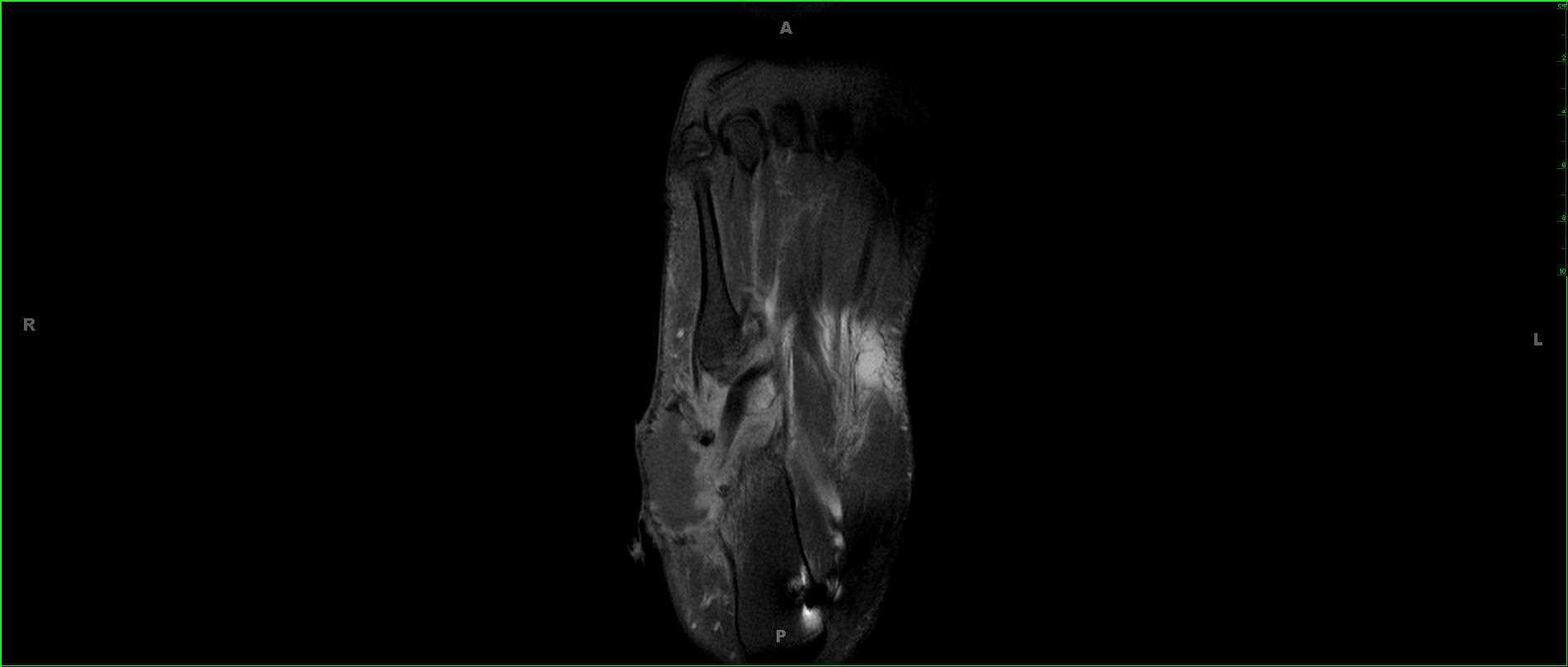
17-year-old male presenting for a painful lump over the lateral aspect of the hindfoot which has been present for several months and slowly enlarging. These images demonstrate a poorly circumscribed T1 hypointense, STIR heterogeneously hyperintense mass centered along the lateral aspect of the hindfoot abutting both the calcaneus, cuboid, and extending towards the base of the fifth metatarsal. The lesion partially envelops the tendons of the peroneus longus and brevis as well as abuts the muscle belly of the abductor digiti minimi. On the postcontrast images, the lesion demonstrates peripheral enhancement with a very poorly circumscribed border. On resection, this lesion was a synovial sarcoma. Synovial sarcomas reflect high-grade malignant soft tissue tumors which typically have an indolent course. Age of presentation is typically 10-20 years with a slight male predominance. Clinical features include a slowly enlarging soft tissue mass with the most common locations including the soft tissues adjacent to large joint. Synovial sarcomas account for approximately 5% of all soft tissue sarcomas. Treatment includes a combination of both surgery and chemoradiation therapy.
Related videos to the case


THIS IS CASE
254
OF
373