- 2
- ,
- 2
- 4
- 6
To Quiz Yourself: Select OFF by clicking the button to hide the diagnosis & additional resources under the case.
Quick Browser: Select ON by clicking the button to hide the additional resources for faster case review.
CASE NUMBER
250


Diagnosis
Ewing Sarcoma, right temporal bone
Note
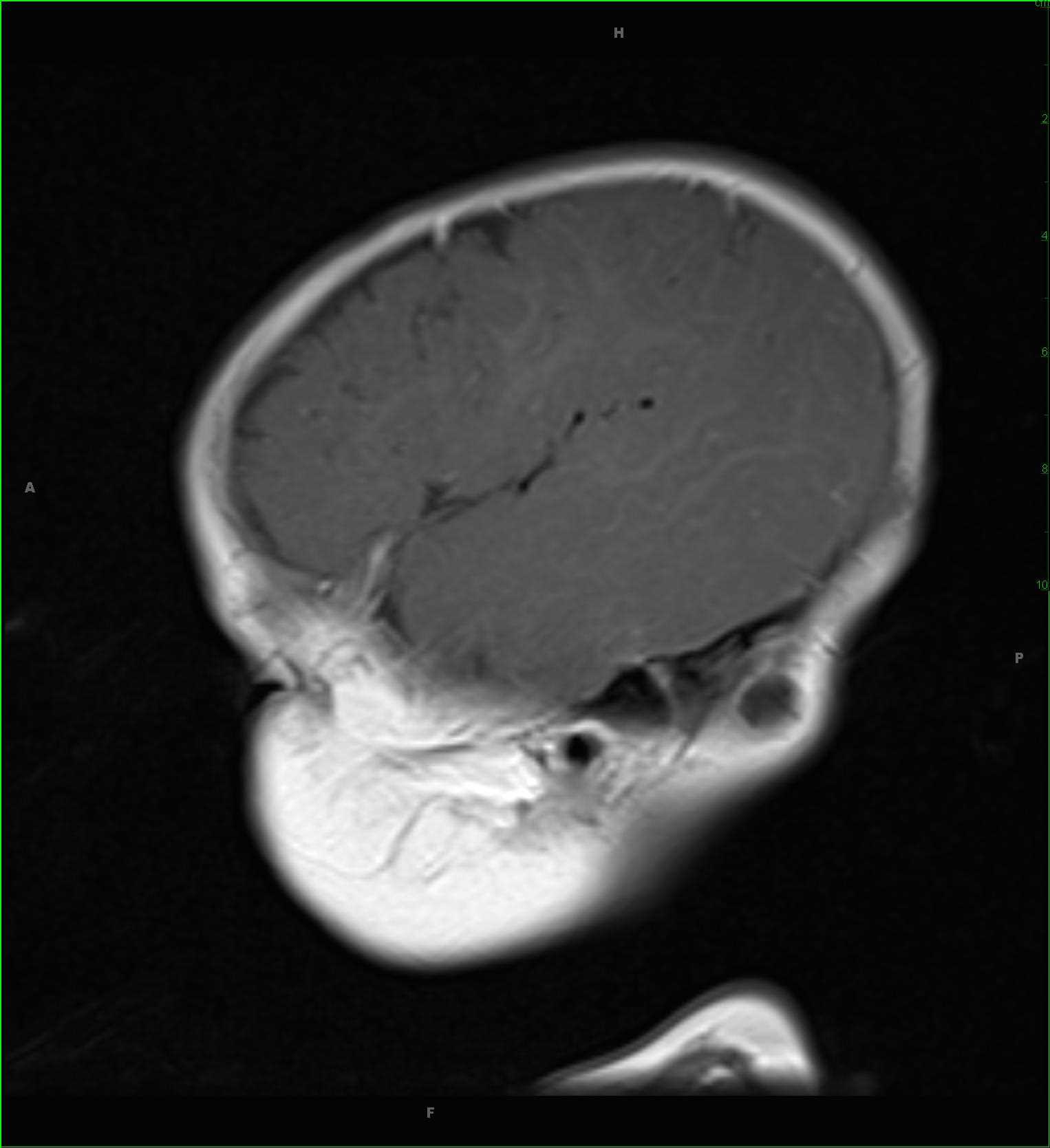
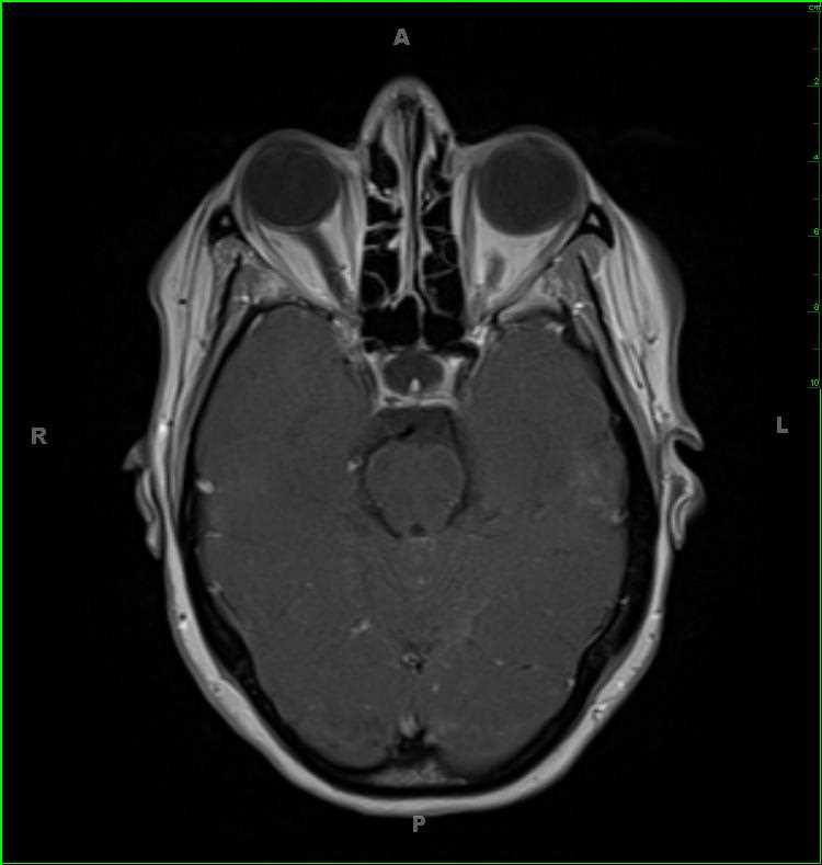
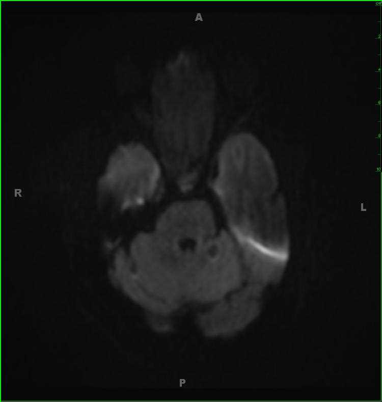
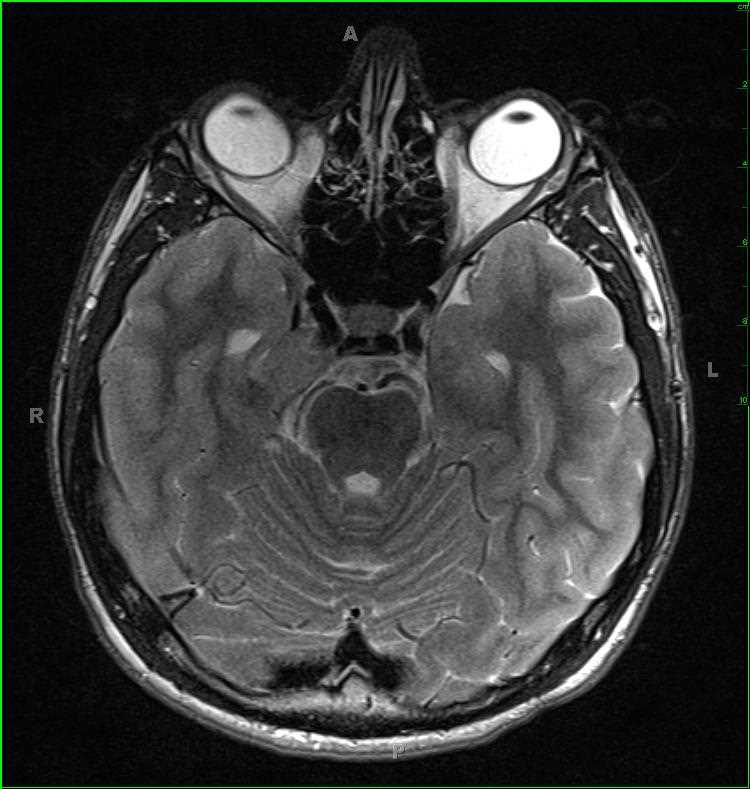
11-year-old male who presented for right facial parasthesias and dysesthesias. On the postcontrast 3D-axial T1-weighted images, there is a round, circumscribed, enhancing, mass with T2/FLAIR mildly hyperintense signal centered within Meckels cave on the right. The lesion is minimally hypointense on the T1-weighted image. Meningioma and schwannoma were given in the differential. On biopsy, this was proven to be an Ewing sarcoma. Ewing sarcoma is the second most common highly malignant primary bone tumor of childhood after osteosarcoma. Typical age at presentation is 10-20 years with a slight male predominance. Pain is the most common presenting symptom. Ewing sarcoma falls into the class of small round blue cell tumors.
Related videos to the case