- 2
- ,
- 2
- 4
- 6
To Quiz Yourself: Select OFF by clicking the button to hide the diagnosis & additional resources under the case.
Quick Browser: Select ON by clicking the button to hide the additional resources for faster case review.
CASE NUMBER
248


Diagnosis
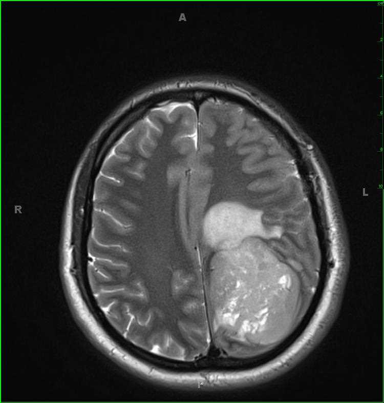
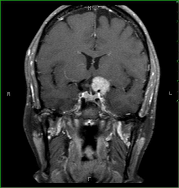
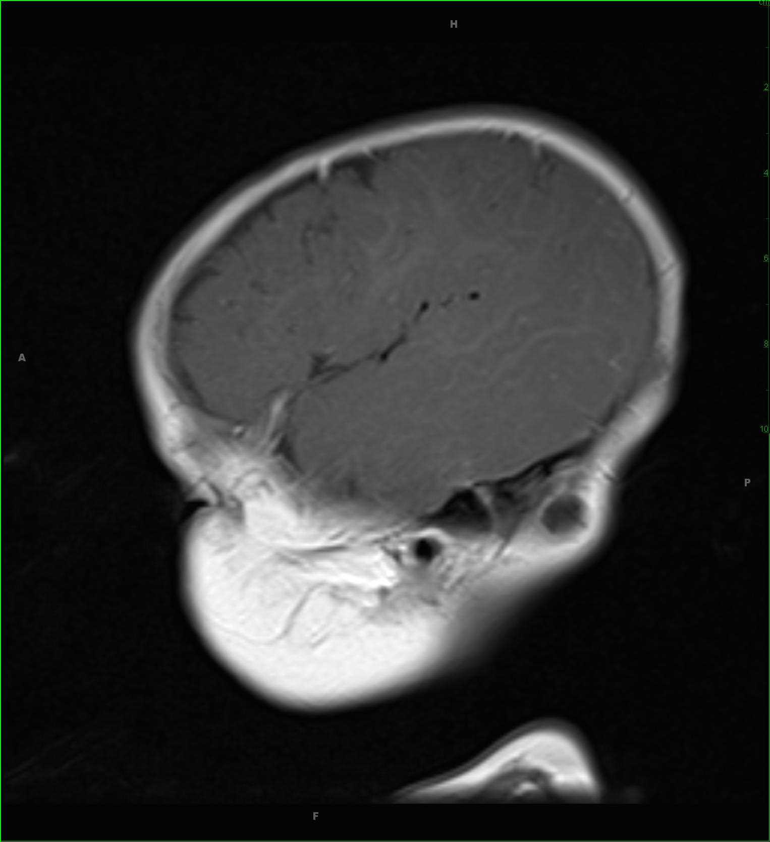
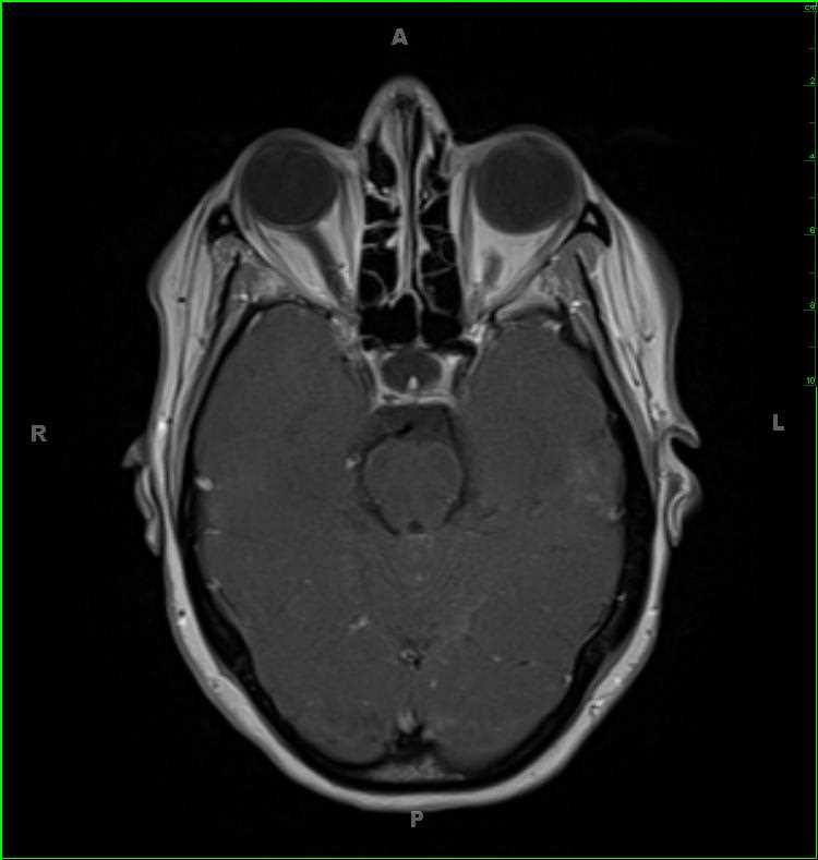
Leptomeningeal Metastatic Disease, lung adenocarcinoma primary
Note
50-year-old male with a primary right upper lobe lung adenocarcinoma. A screening MRI of the brain was ordered for evaluation for metastatic disease. There are scattered, linear regions of minimal leptomeningeal thickening and enhancement within the supratentorial compartment. There is a more nodular focus of abnormal T2/FLAIR-hyperintense signal in the right central gyrus with corresponding enhancement. More inferiorly, over the superior vermis and cerebellar hemispheres, there is also thickening and enhancement of the leptomeninges. The final image demonstrates thickening and enhancement of the cisternal and canalicular segments of cranial nerves VII and VIII. The differential includes meningitis, hemorrhage, carcinomatosis, intracranial hypotension, vasculitis and granulomatous inflammation. Given the stated clinical history, these findings are compatible with leptomeningeal spread of disease.
Related videos to the case