- 2
- ,
- 2
- 4
- 6
To Quiz Yourself: Select OFF by clicking the button to hide the diagnosis & additional resources under the case.
Quick Browser: Select ON by clicking the button to hide the additional resources for faster case review.
CASE NUMBER
241


Diagnosis
Pituicytoma
Note
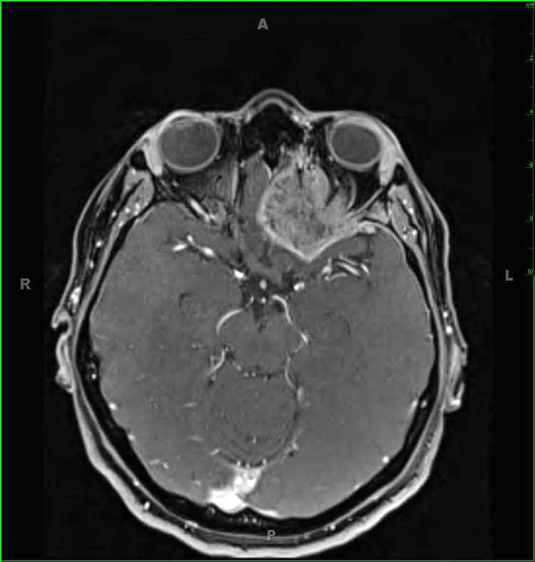
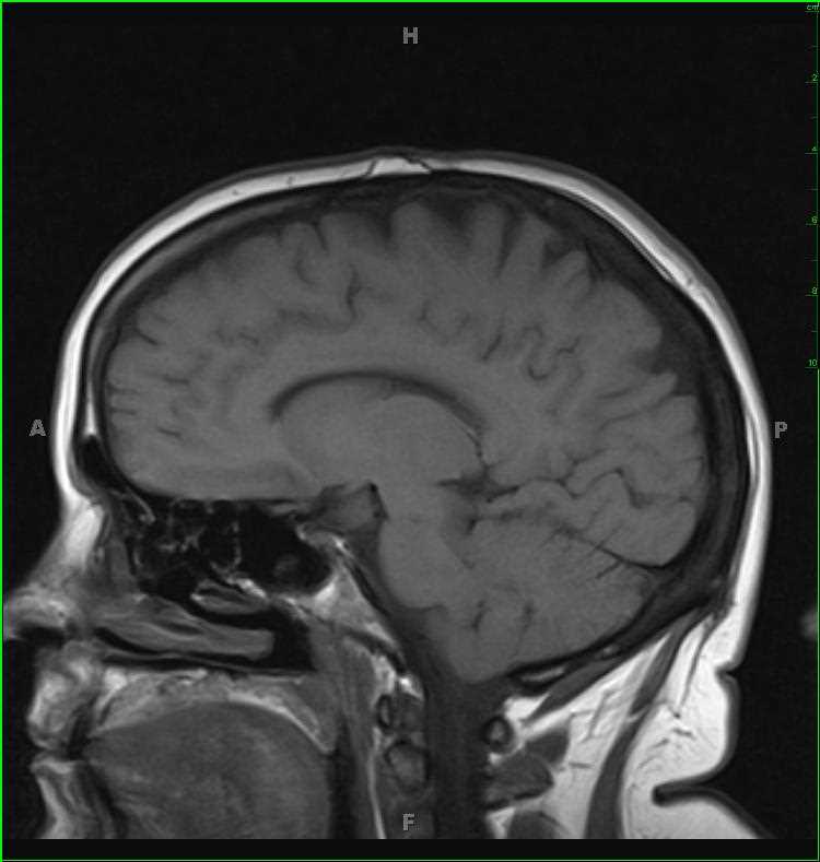
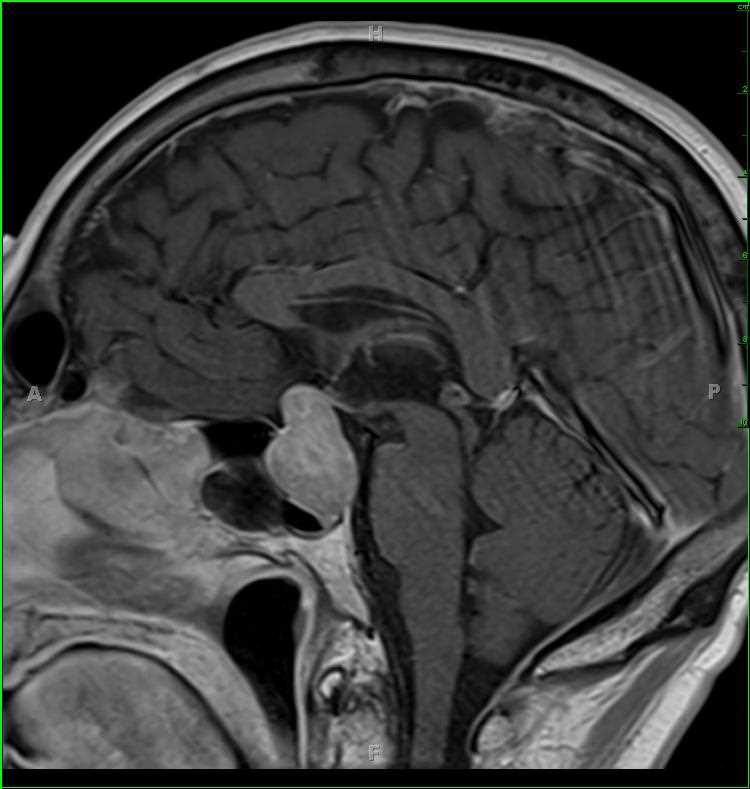
51-year-old male with a history of pan hypopituitarism. There is a well circumscribed, solid appearing T1-slightly hypointense, T2/FLAIR mildly hyperintense mass with diffusion restriction and enhancement. The lesion enlarges the sella turcica with chronic remodeling/erosion of the posterior wall of the sella turcica. There is elevation of the infindibulum, no cavernous sinus invasion, and contact with and superior displacement and deformity of the optic chiasm and prechiasmatic optic nerve segments. The differential diagnosis includes pituitary macroadenoma, pituicytoma, granular cell tumor of the pituitary gland and astrocytoma of the neurohypophysis among others. This was a biopsy proven pituicytoma. Pituicytomas arise from pituicytes which are specialized glial cells in the neurohypophysis and infundibulum. The peak incidence is in the 5th decade, with a female to male ratio of 2:1. Clinical presentation is either from endocrine dysfunction or from compression of adjacent structures.
Related videos to the case