- 2
- ,
- 2
- 4
- 6
To Quiz Yourself: Select OFF by clicking the button to hide the diagnosis & additional resources under the case.
Quick Browser: Select ON by clicking the button to hide the additional resources for faster case review.
CASE NUMBER
198


Diagnosis
Spinal Cord Infarct, secondary to vertebral artery dissection
Note
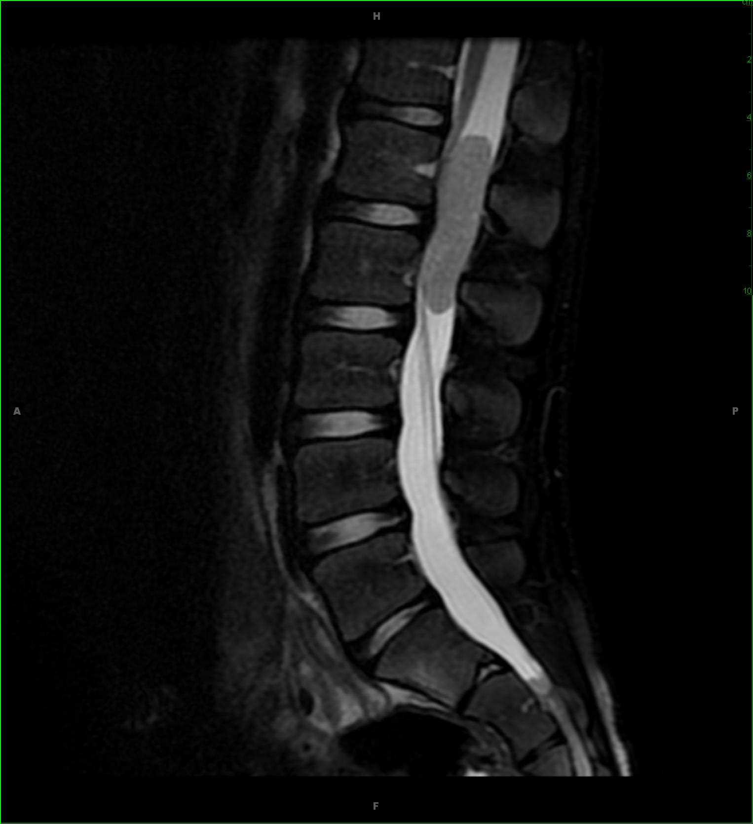
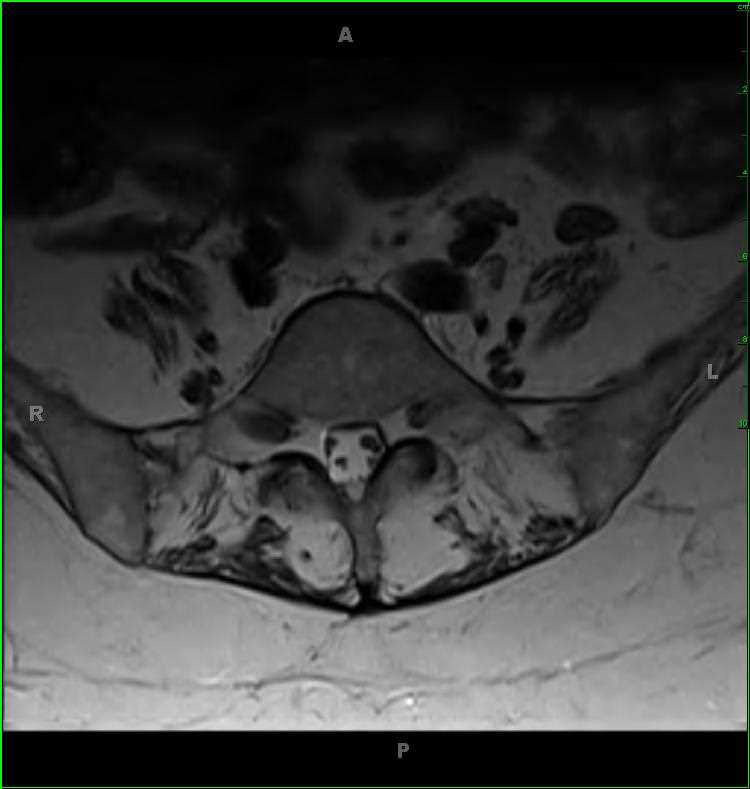
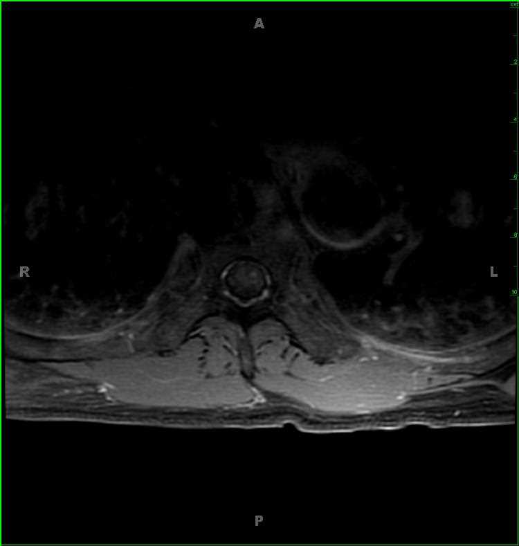
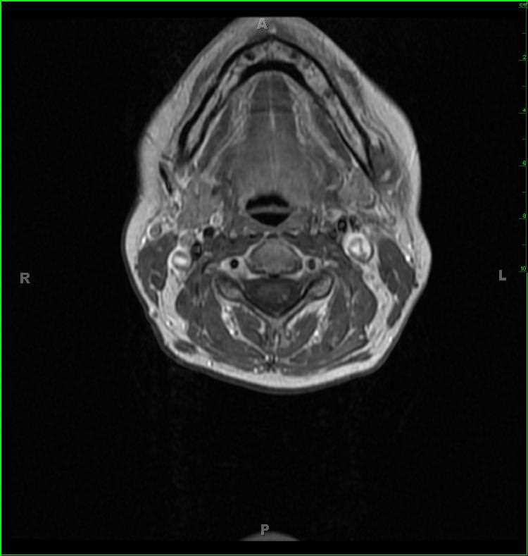
36-year-old female with sudden onset left upper extremity parasthesias and weakness. There is a well circumscribed, ovoid, T1 slightly hyperintense, T2/STIR-hyperintense lesion within the left posterior aspect of the upper cervical spinal cord. The lesion has convex borders and enhances. Differential diagnosis includes infectious/inflammatory demyelinating focus, underlying vascular malformation, glioma or metastatic disease. On further imaging, multiple acute infarcts were identified in the left anterior inferior and posterior inferior cerebral artery territories. A subsequent 4-vessel angiogram demonstrated a dissection within the distal second segment of the left vertebral artery. The diagnosis for the cervical spine signal abnormality in this case was a focal infarct. Most cases of infarct are idiopathic with atherosclerosis attributed as the cause in approximately 1/3 of cases. Other etiologies include trauma, embolism, hypotension, and radiation induced vasculopathy among many others. Symptoms appear quickly, with maximum symptomatology at 12 hours post ictus.
Related videos to the case