- 2
- ,
- 2
- 4
- 6
To Quiz Yourself: Select OFF by clicking the button to hide the diagnosis & additional resources under the case.
Quick Browser: Select ON by clicking the button to hide the additional resources for faster case review.
CASE NUMBER
194


Diagnosis
Spinal Cord Ependymoma
Note
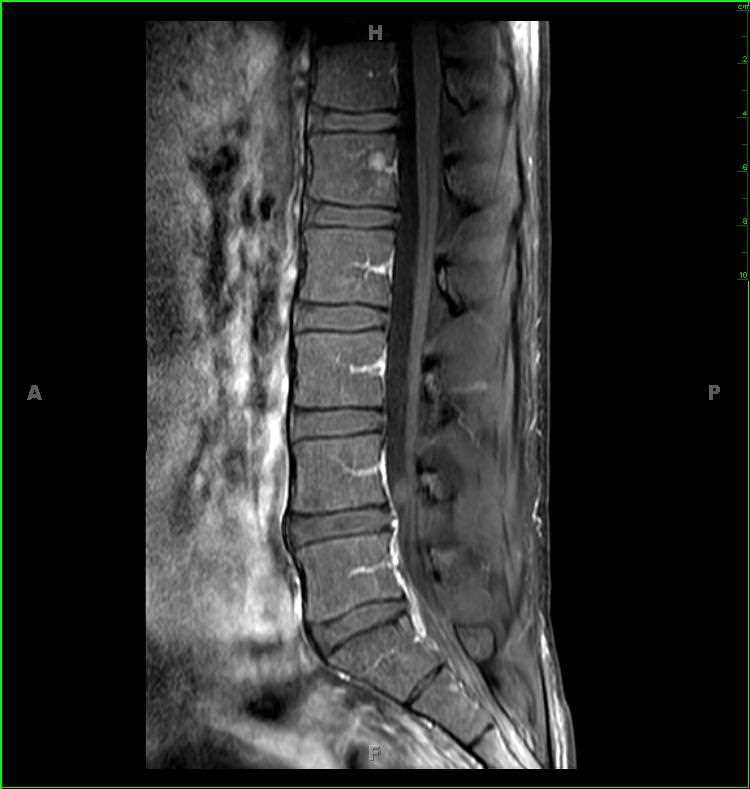
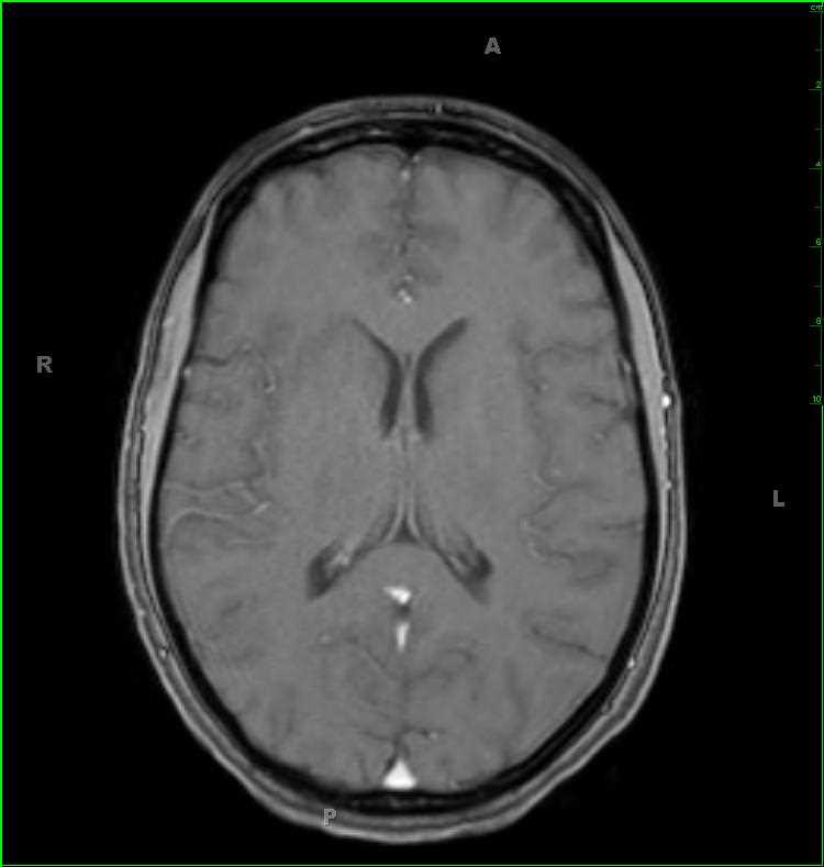
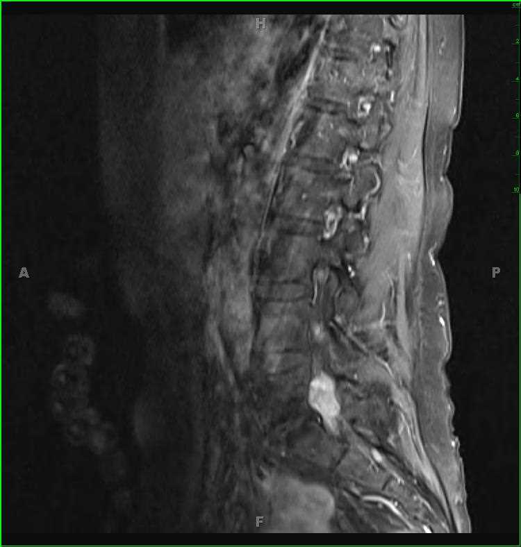
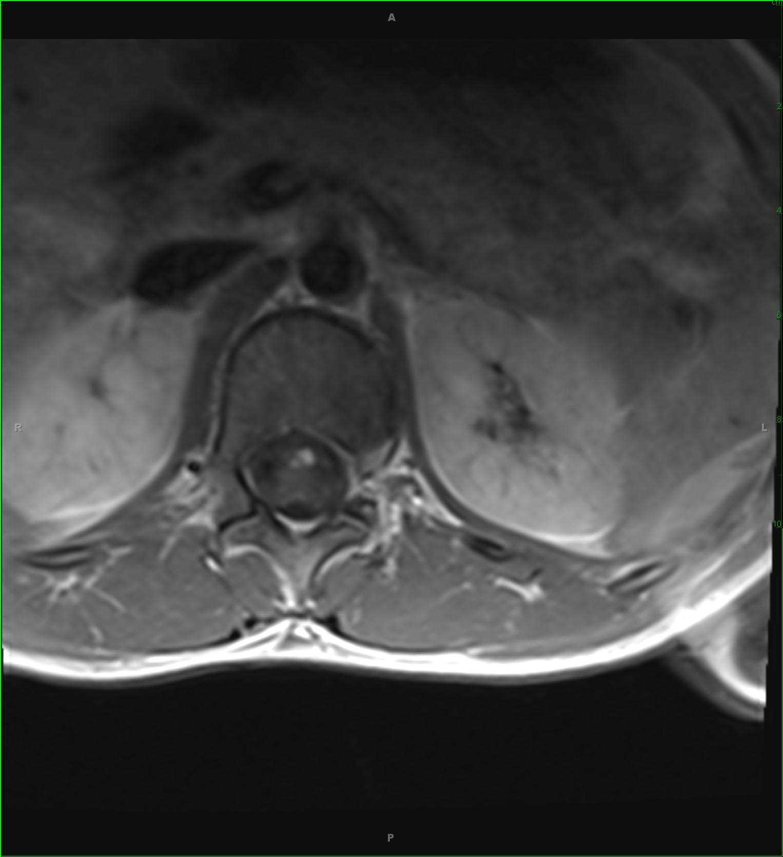
11-year-old female with a history of scoliosis. The patient was previously evaluated with a noncontrast spinal MRI which demonstrated a large central region of T2-hyperintense and T1-hypontense signal within the central cord substance from the lower cervical to upper thoracic spinal region suspicious for syringohydromyelia. There was no Chiari malformation. The patient was brought back for postcontrast images which revealed an infiltrative mass with nodular features within the central cord substance, greatest in the inferior thoracic spinal region. There are cystic components of the lesion at its cranial and caudalmost portions. In addition, there are regions of nodular enhancement along the ventral aspect of the conus medullaris compatible with leptomeningeal metastatic disease. The differential diagnosis for the imaging findings includes astrocytoma, ependymoma, and hemangioblastoma. This was a case of cord ependymoma. Intramedullary spinal cord ependymomas are rare, accounting for only 4-10% of all CNS neoplasms. Spinal cord ependymomas are more common in adults, while astrocytomas are more common in children.
Related videos to the case


THIS IS CASE
194
OF
373