- 2
- ,
- 2
- 4
- 6
To Quiz Yourself: Select OFF by clicking the button to hide the diagnosis & additional resources under the case.
Quick Browser: Select ON by clicking the button to hide the additional resources for faster case review.
CASE NUMBER
163


Diagnosis
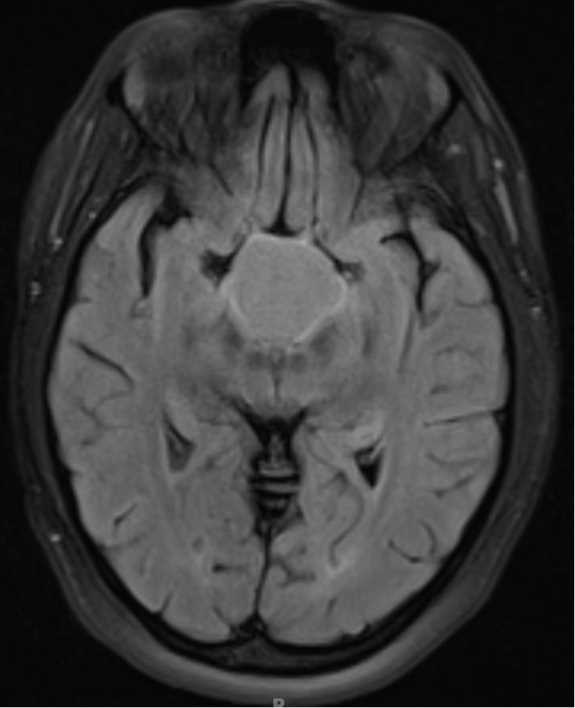
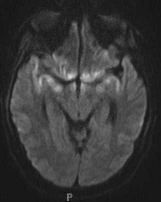
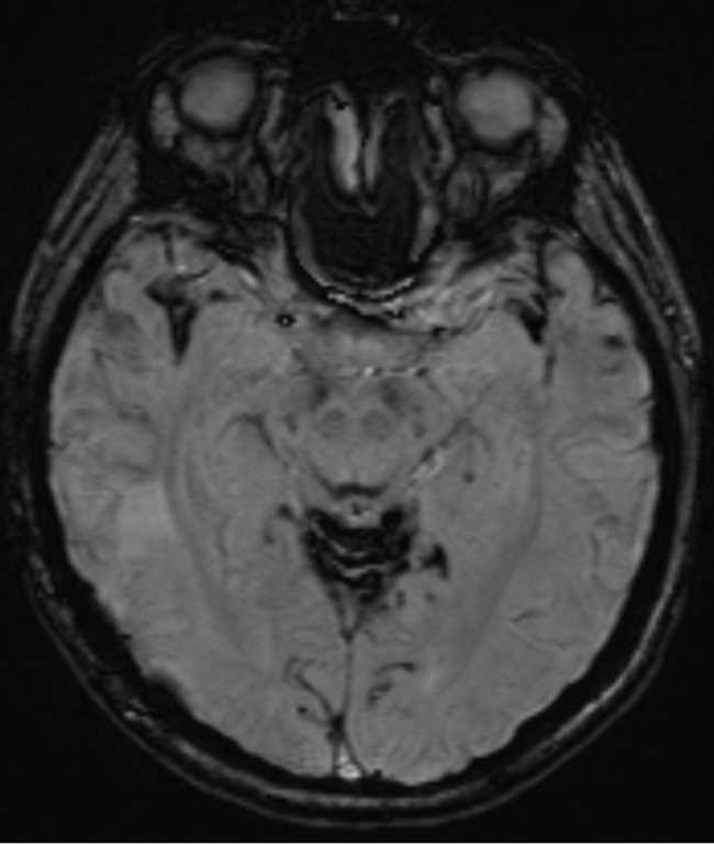
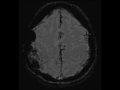
Superficial Siderosis
Note
This is a case of superficial siderosis in a patient with a history of a right parietal AVM status post resection. A resection cavity with associated gliotic margins is demonstrated. On the susceptibility weighted images, there are numerous sights of curvilinear signal loss within numerous sulci of the supratentorial compartment. There is involvement of the superior folia of the vermis and cerebellar hemispheres. There is also involvement of the margins of the resection cavity. Superficial siderosis results from recurrent episodes of subarachnoid hemorrhage resulting in hemosiderin deposition on the surface of the brain, brainstem, and cranial nerve leptomeningeal surfaces. Common causes of superficial siderosis include trauma, vascular malformations and aneurysms, hemorrhagic CNS neoplasms and previous surgery.
Related videos to the case

THIS IS CASE
163
OF
373