- 2
- ,
- 2
- 4
- 6
To Quiz Yourself: Select OFF by clicking the button to hide the diagnosis & additional resources under the case.
Quick Browser: Select ON by clicking the button to hide the additional resources for faster case review.
CASE NUMBER
155


Diagnosis
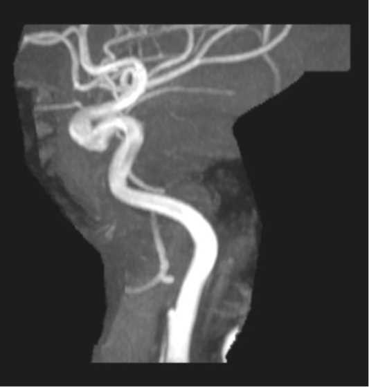
Cervical ICA Dissection and Pseudoaneurysm
Note
On the 3-D time of flight MRA images of the head, there is a small dissection flap arising from the anterior wall of the cervical ICA segment on the right. More superiorly, there is a saccular outpouching compatible with a pseudoaneurysm at that site. The 3-D MIP images redemonstrate the saccular pseudoaneurysm. Pseudoaneurysms characteristically lack all three layers of the arterial wall (intima, media, and adventitia). Pseudoaneurysm development may occur at any time point following initial arterial injury. Most cases will present within 5 years of the injury. Etiologies include trauma, dissection, vasculitides, infection or iatrogenic causes.
Related videos to the case