- 2
- ,
- 2
- 4
- 6
To Quiz Yourself: Select OFF by clicking the button to hide the diagnosis & additional resources under the case.
Quick Browser: Select ON by clicking the button to hide the additional resources for faster case review.
CASE NUMBER
151


Diagnosis
Lumbar Insufficiency Fracture
Note
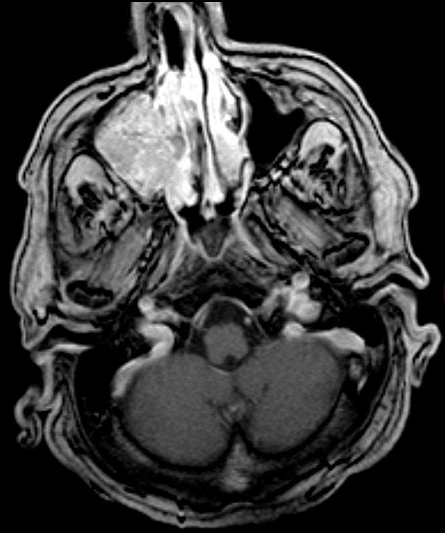
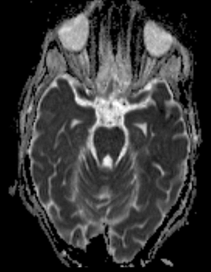
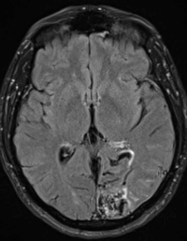
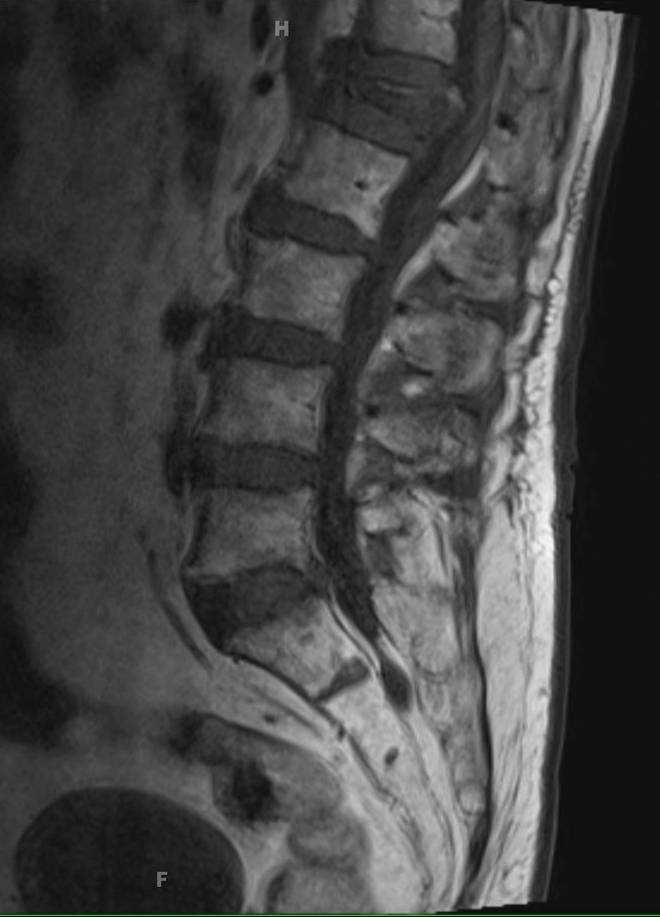
68-year-old female who presented for worsening back pain over 2 weeks. The sagittal T1 weighted image demonstrates moderate superior and minimal inferior compression deformities of the L1 vertebral body, with loss of approximately 50% vertebral body height anteriorly. There is diffuse T1 hypointense signal in the L1 vertebral body. On the sagittal T2 weighted images, there is mild retropulsion of osseous fragments into the spinal canal resulting in partial effacement of the thecal sac with contact and mild deformity of the ventral aspect of the conus medullaris. The sagittal STIR image, where there is also demonstration of diffuse marrow edema. The axial T2 and postcontrast axial T1 weighted images also demonstrate the partially effaced thecal sac. On the postcontrast sagittal T1 weighted image, there are no suspicious regions of enhancement within the marrow space of vertebra L1. This is a case of a vertebral insufficiency fracture which results from axial loading on osteoporotic bone with lost of anterior vertebral body height. In the acute setting, there is low T1 signal with high T2 and STIR signal due to edema with gradual normalization over time. 50-65% of traumatic vertebral compression fractures are between T12 and L2, but osteoporotic compression fractures can occur at any level. The superior endplate is most commonly involved.
Related videos to the case