- 2
- ,
- 3
- 2
- 7
To Quiz Yourself: Select OFF by clicking the button to hide the diagnosis & additional resources under the case.
Quick Browser: Select ON by clicking the button to hide the additional resources for faster case review.
CASE NUMBER
323


Diagnosis
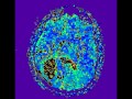
Intracranial Abscess
Note
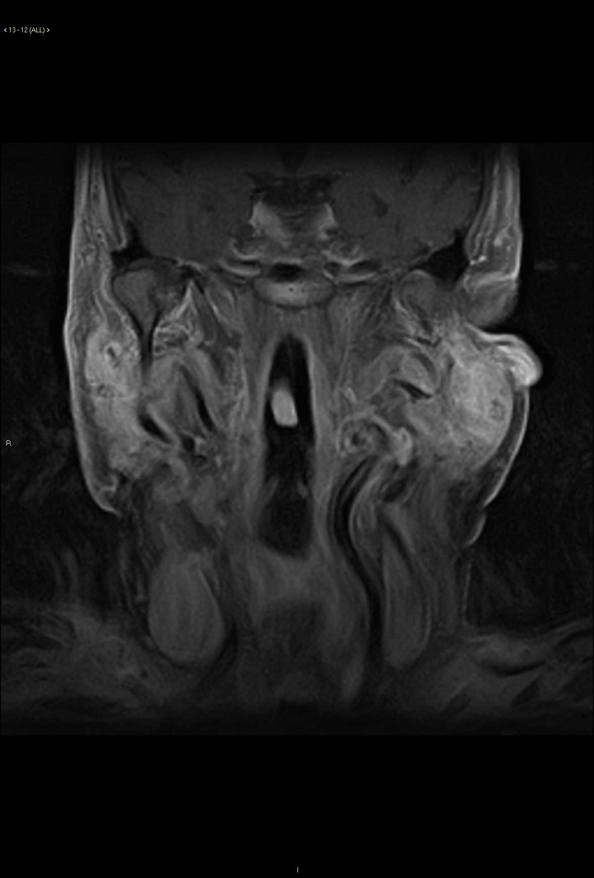
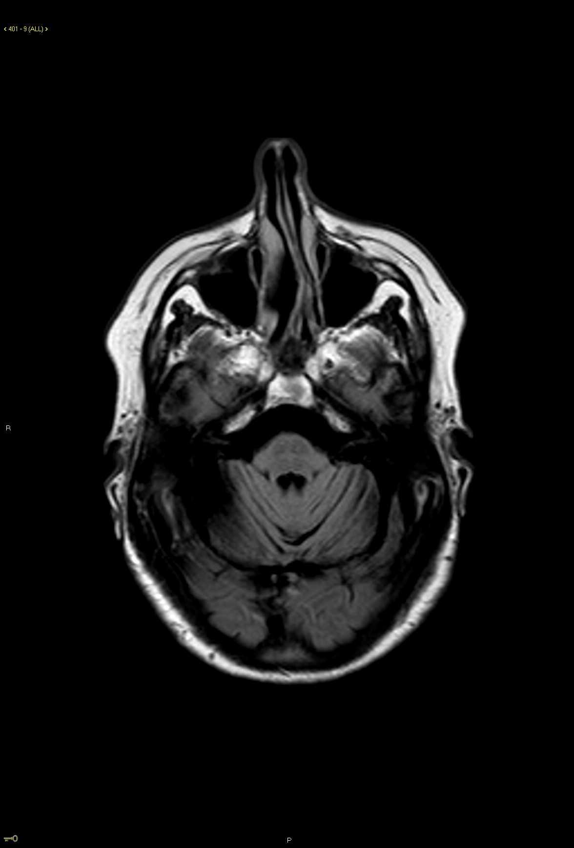
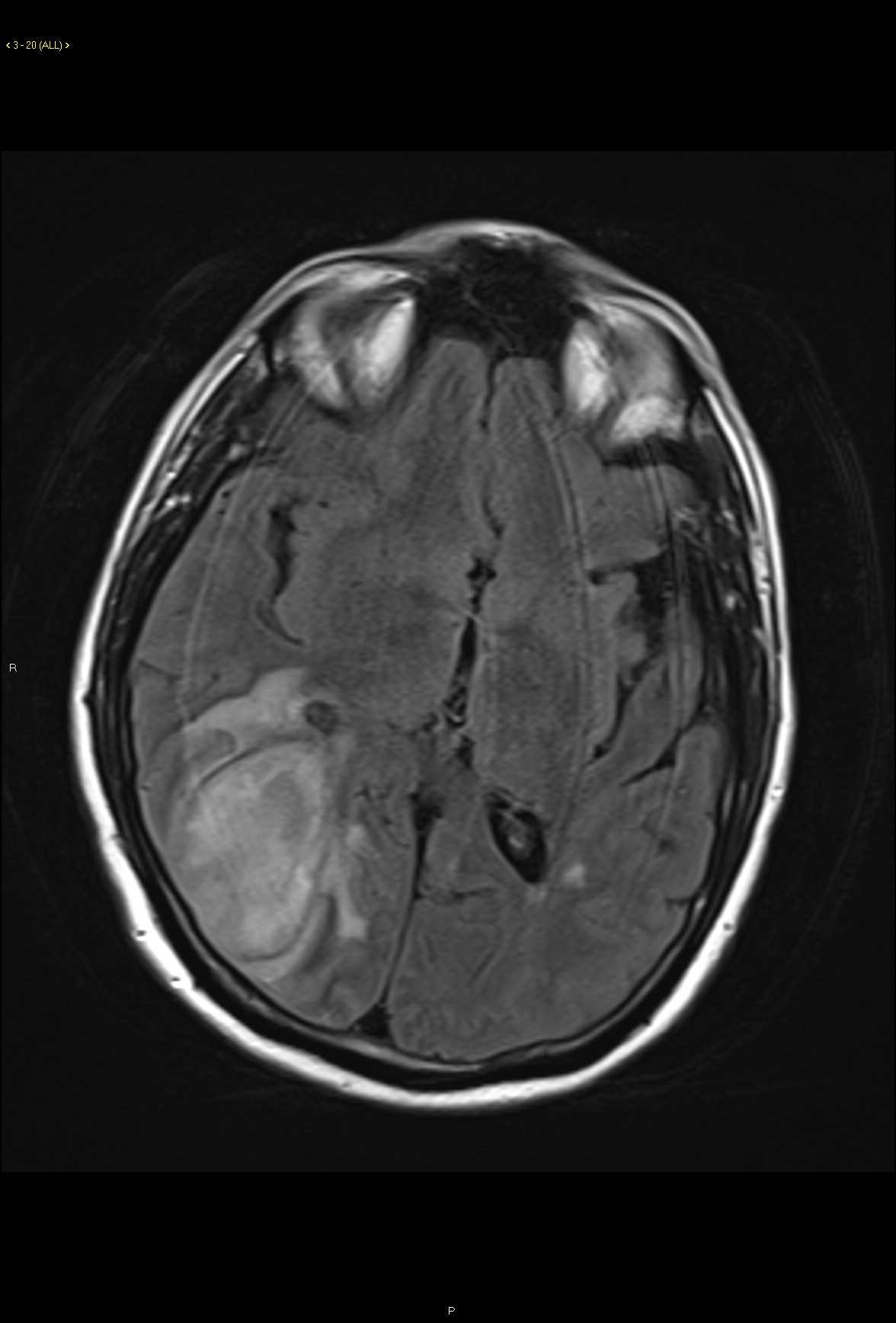
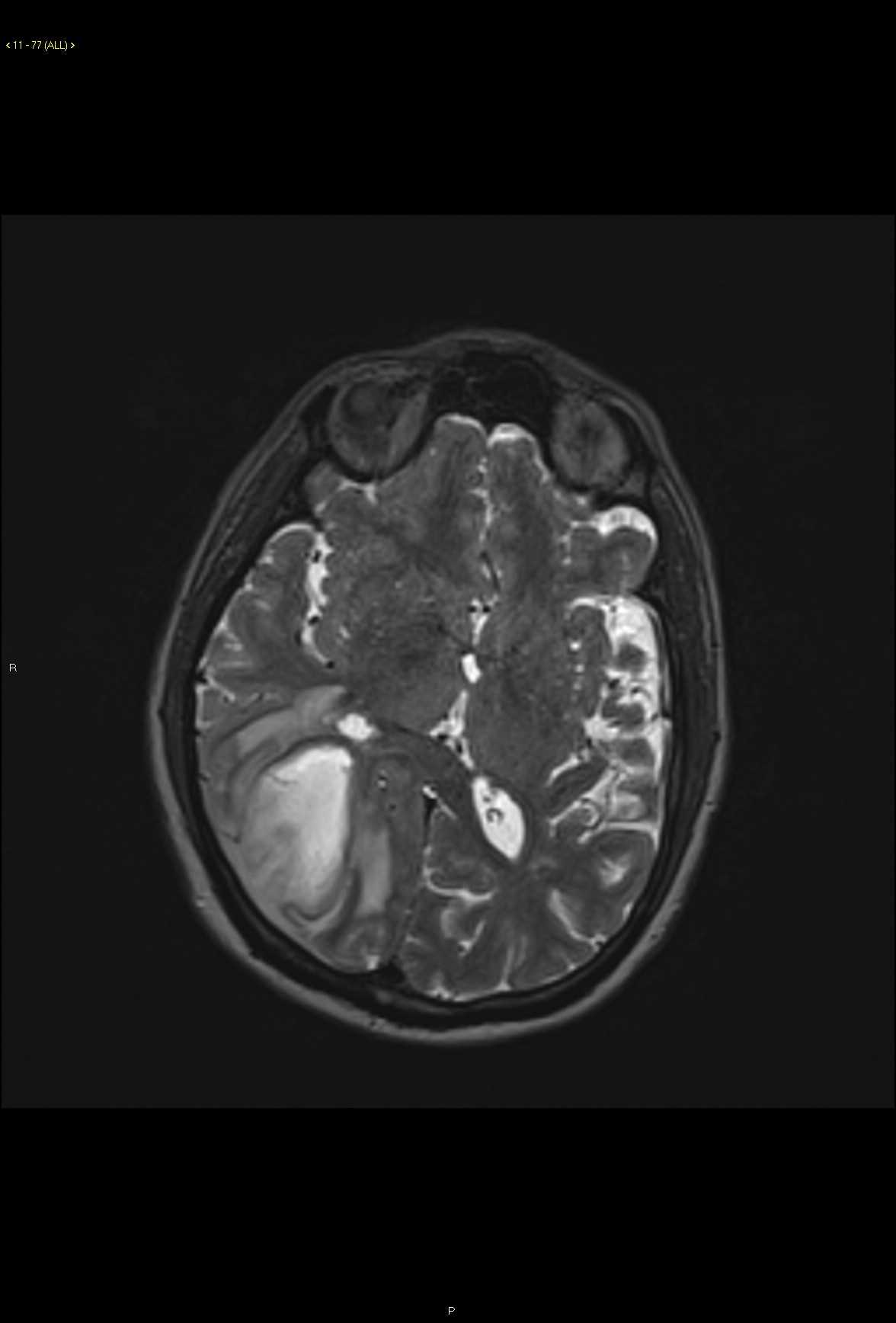
56-year-old male with new onset seizures. There is a circumscribed T2/FLAIR hyperintense mass centered within the right occipitoparietal region. There is significant perilesional edema and effacement of the surrounding cerebral sulci. On the diffusion weighted images, there is a central region of diffusion restriction within the lesion with spillage of diffusion restricting contents into the adjacent subdural space over the posterior right cerebral hemisphere. There is scattered susceptibility artifact along the margins of the lesion extending to the subdural space with peripheral hyperemia on the perfusion weighted images. The lesion demonstrates an irregular rind of enhancement on the postcontrast images. Findings are most compatible with a brain parenchymal abscess with an associated subdural abscess. Four distinct stages in the development or evolution of a brain abscess consisting of early cerebritis, late cerebritis, early capsule and late capsule formation.
Related videos to the case

THIS IS CASE
323
OF
370