- 2
- ,
- 3
- 2
- 7
To Quiz Yourself: Select OFF by clicking the button to hide the diagnosis & additional resources under the case.
Quick Browser: Select ON by clicking the button to hide the additional resources for faster case review.
CASE NUMBER
321


Diagnosis
Carcinoma ex pleomorphic adenoma
Note
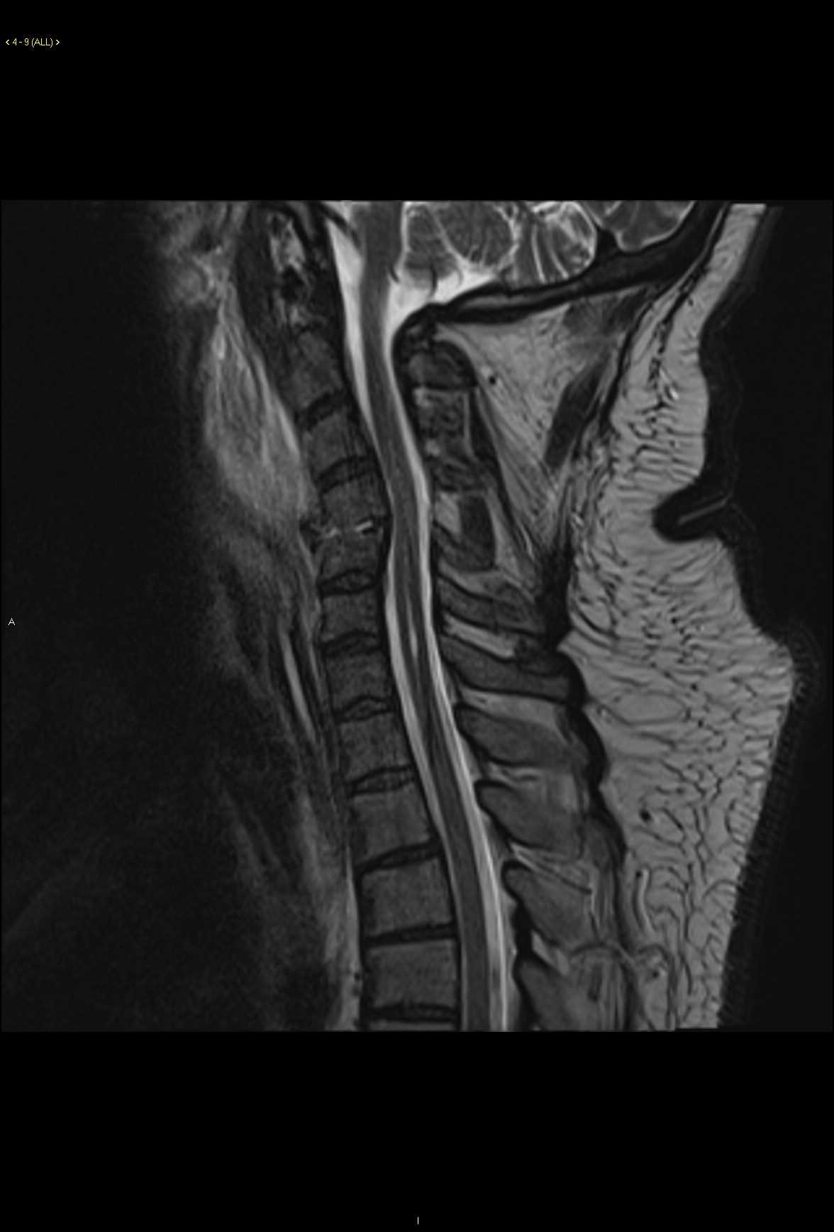
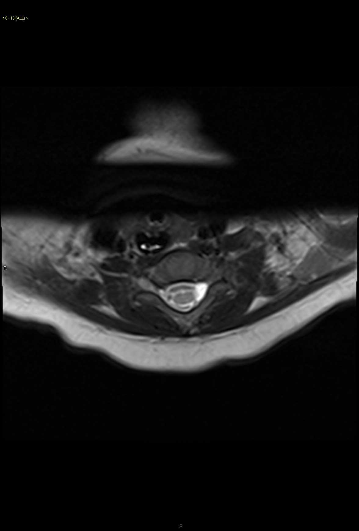
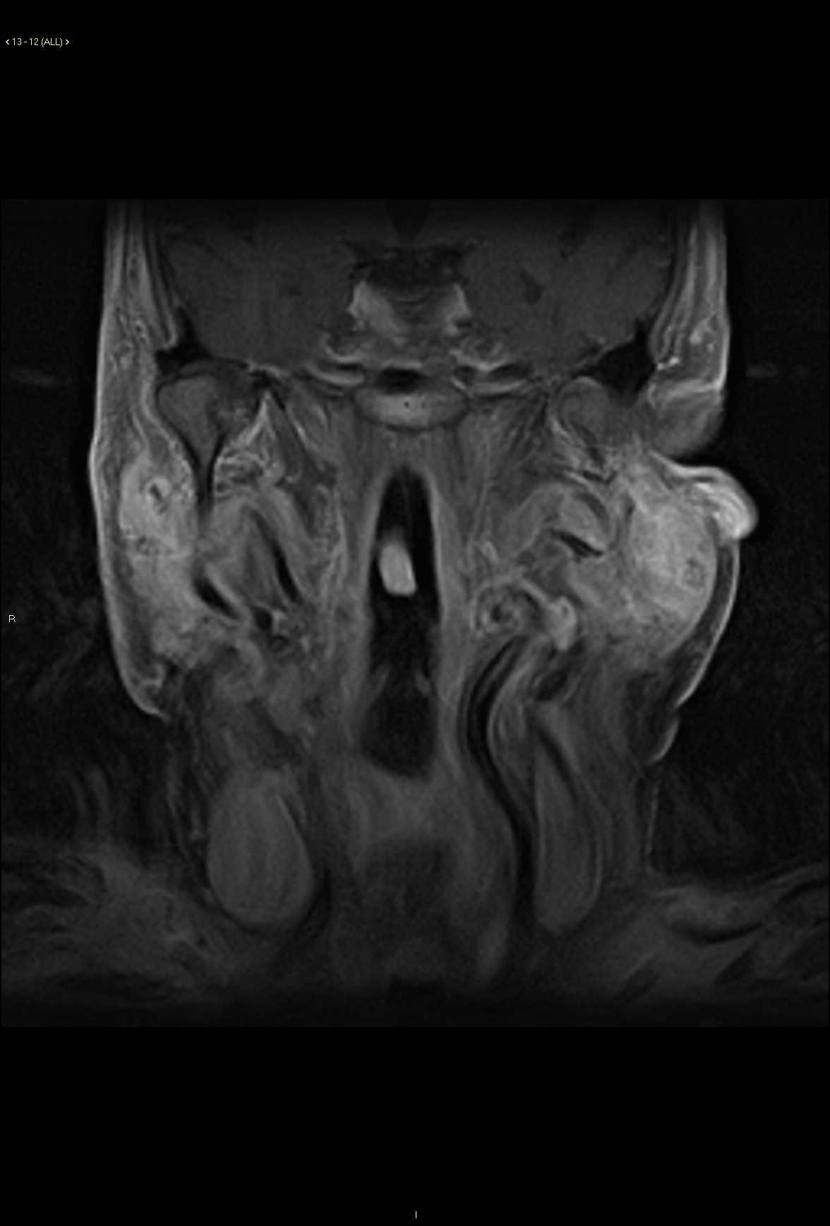
54-year-old female with a history of a slowly enlarging left facial mass. There is a circumscribed solid cystic T1 hypointense, T2/STIR heterogeneously hyperintense mass centered within the superficial left parotid gland. The lesion demonstrates heterogeneous enhancement with scattered nonenhancing cystic or necrotic regions. A differential diagnosis of pleomorphic adenoma, lymphoma, and metastatic disease was given. Carcinoma ex pleomorphic adenoma was found on biopsy. Carcinoma ex pleomorphic adenoma is malignant mixed tumor of the parotid. The other subtype of malignant mixed tumor is carcinosarcoma. Small carcinoma ex pleomorphic adenoma have a similar imaging appearance to pleomorphic adenoma. Larger lesions tend to invade surrounding structures. A history of a rapidly enlarging parotid mass is a concerning feature. Other symptoms may be present such as facial weakness.
Related videos to the case


THIS IS CASE
321
OF
370