- 2
- ,
- 3
- 2
- 7
To Quiz Yourself: Select OFF by clicking the button to hide the diagnosis & additional resources under the case.
Quick Browser: Select ON by clicking the button to hide the additional resources for faster case review.
CASE NUMBER
317


Diagnosis
Schwannoma
Note
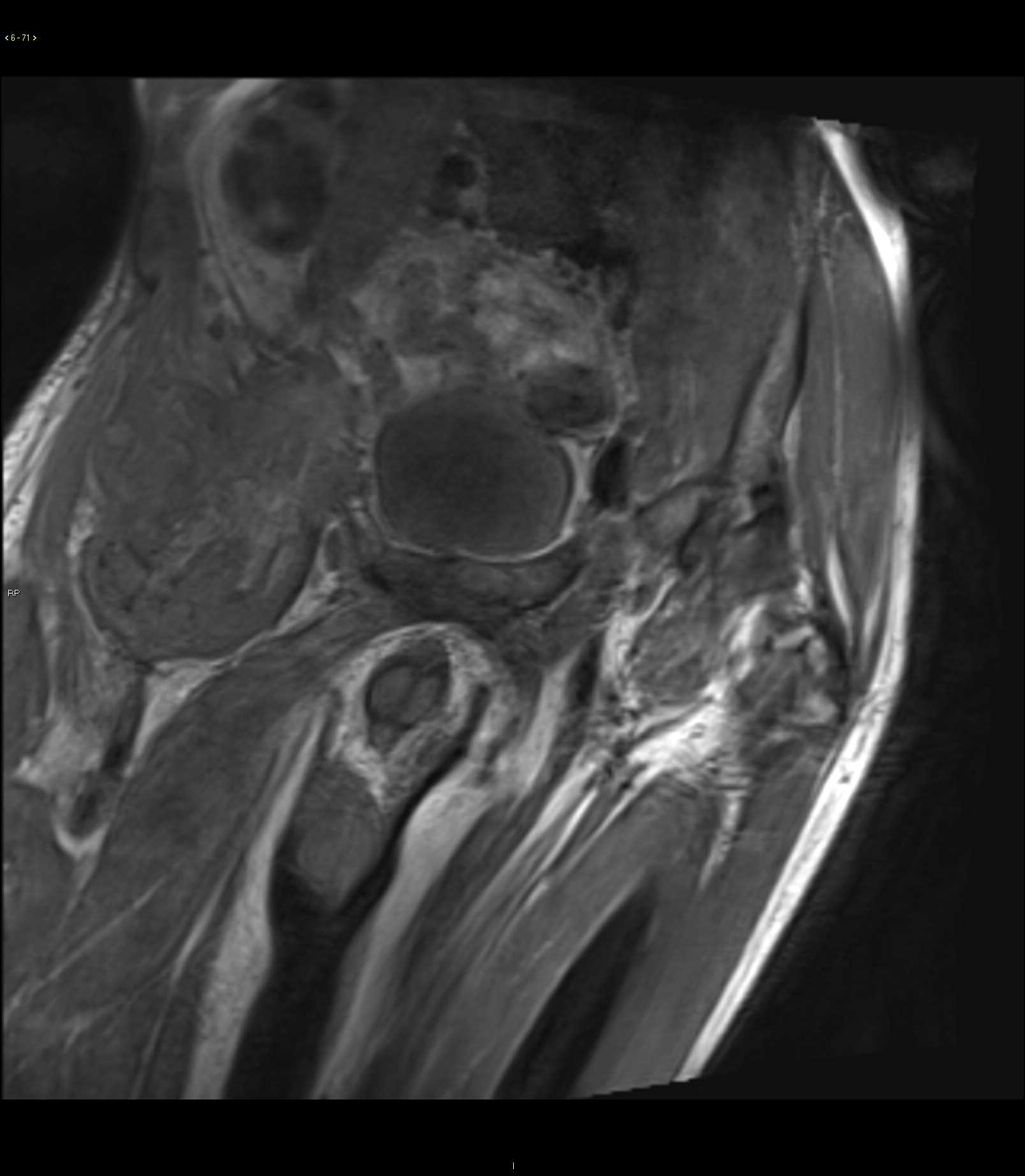
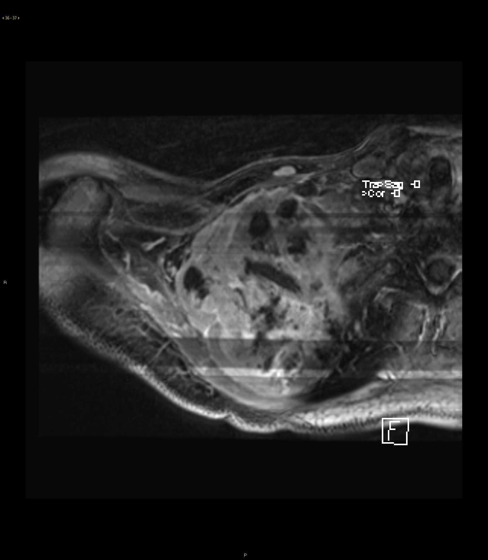
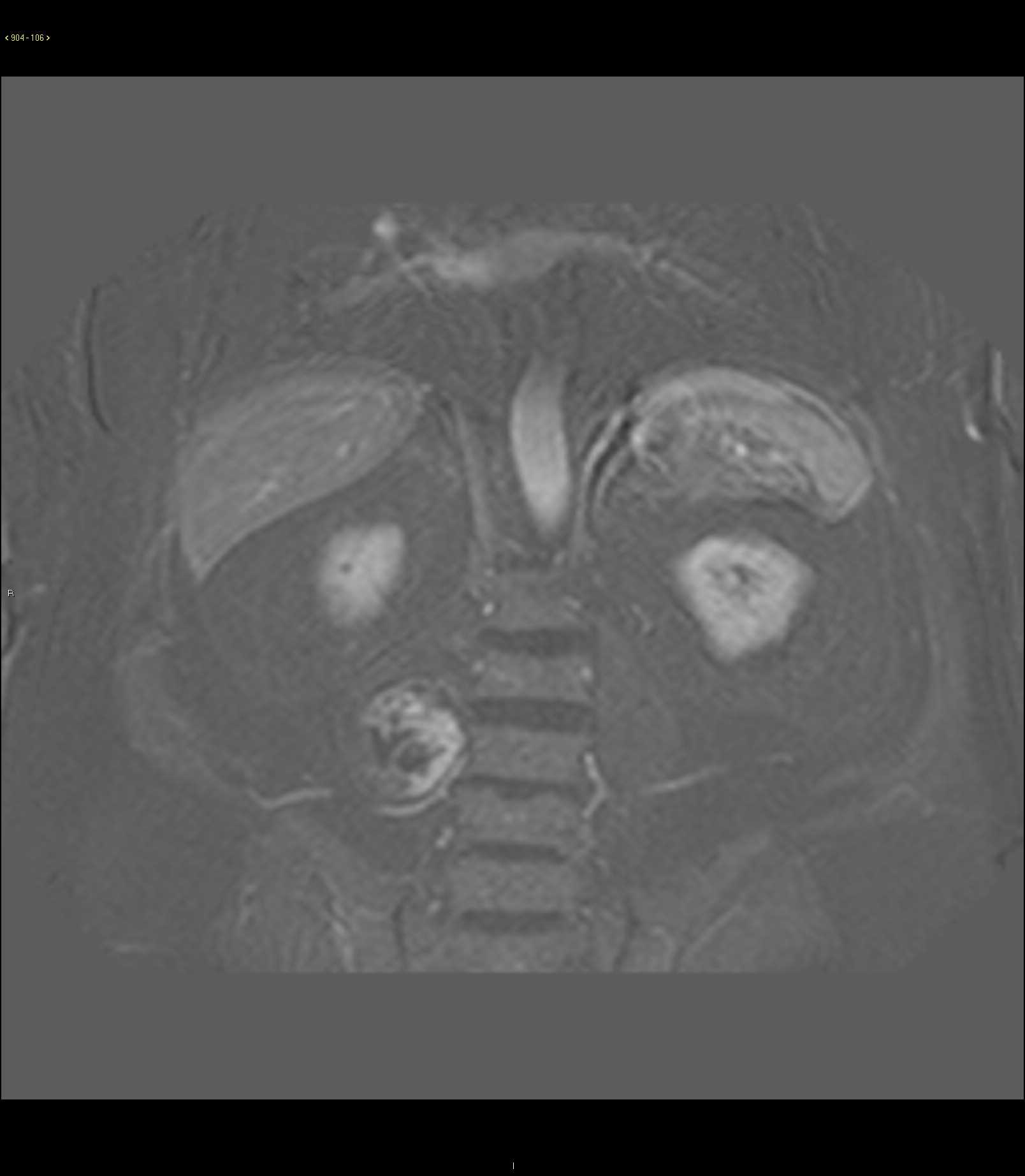
34-year-old male who underwent screening upper abdominal MRI with and without contrast demonstrating a fusiform shaped T1 mildly heterogeneous T2 moderately heterogeneous lesion with both hyper- and hypointense components arising from the L2 nerve root on the right with extension towards the L2-L3 neural foramen. There is evidence of subtle scalloping of the far lateral aspect of the L3 vertebral body. Imaging findings and location are most compatible with an incidentally detected peripheral nerve sheath tumor. Peripheral nerve sheath tumors can be divided into 2 major benign categories: neurofibroma and schwannoma. Malignant category is represented by the malignant peripheral nerve sheath tumor. Neurofibromas are most common in the setting of neurofibromatosis type I with schwannomas present in the setting of neurofibromatosis type II or systemic schwannomatosis. The important imaging features to evaluate for the possibility of malignant degeneration includes the presence of restricted diffusion, early arterial phase enhancement, and interval growth in comparison to prior studies.
Related videos to the case